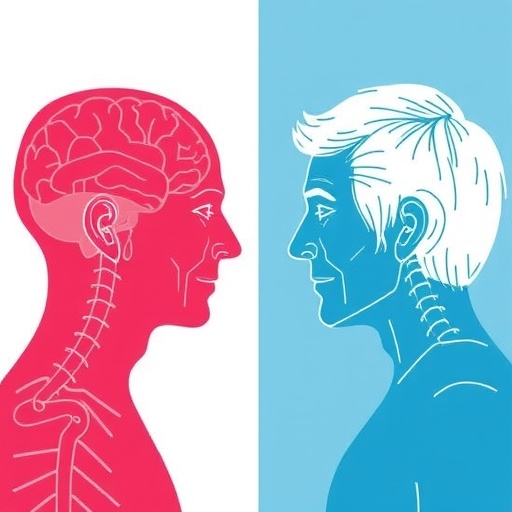
In a groundbreaking development poised to revolutionize Parkinson’s disease diagnostics, an international team of researchers has unveiled a novel imaging analysis approach that distinguishes between two hypothesized subtypes of the disease: Brain-First and Body-First Parkinson’s. This advancement stems from combining conventional dopamine transporter (DAT) SPECT imaging techniques with sophisticated radiomics-enhanced data analytics, potentially illuminating the elusive origins and progression pathways of Parkinson’s disease (PD) in unprecedented detail.
Parkinson’s disease, a debilitating neurodegenerative disorder, affects millions worldwide, characterized by the loss of dopamine-producing neurons leading to motor dysfunction and a spectrum of non-motor symptoms. Despite decades of research, teasing apart the heterogeneous nature of PD has remained a foremost challenge. The dichotomous theory posits that some patients experience an initial pathological insult in the brain (Brain-First subtype), while in others, the disease process begins in the peripheral autonomic nervous system before ascending to the brain (Body-First subtype). This crucial differentiation could tailor early intervention strategies, but has been limited by a lack of definitive biomarkers to reliably classify patients.
This new study, appearing in the prestigious journal npj Parkinson’s Disease, leverages the well-established technique of dopamine transporter single-photon emission computed tomography (DAT-SPECT). DAT-SPECT is routinely employed to visualize striatal dopaminergic integrity, serving as a surrogate marker for neurodegeneration in PD. However, solely relying on conventional DAT-SPECT metrics has not been sufficient to disentangle the proposed Brain-First versus Body-First subtypes. The innovation arises by integrating radiomics, an emerging field that extracts a large number of quantitative features from medical images using advanced computational algorithms.
Radiomics can unveil subtle patterns and textures within images imperceptible to the human eye and standard metrics. By applying radiomics-enhanced analysis to DAT-SPECT brain scans, the research team identified distinguishing signatures correlated with the two PD subtypes. These signatures capture nuanced heterogeneity in tracer uptake distribution, asymmetry, and shape characteristics of the striatal dopaminergic deficit. Such imaging phenotypes open avenues to classify individual patients more precisely and understand the underlying pathological geography.
Importantly, the study recruited a rigorously phenotyped patient cohort, encompassing newly diagnosed PD individuals along the suggested Brain-First and Body-First trajectories. The researchers validated their radiomics-based classification model against conventional visual and semi-quantitative assessments, demonstrating superior performance in discriminating subtypes. This breakthrough underscores the potential of combining conventional nuclear imaging with high-dimensional radiomic feature extraction to enhance diagnostic granularity, paving the way for personalized medicine in Parkinson’s.
Delving into the methodology, raw DAT-SPECT images underwent meticulous preprocessing to standardize spatial and intensity parameters, ensuring robustness across multi-center datasets. From the processed images, over one hundred radiomic features were extracted encompassing first-order statistics, shape, texture, and intensity-based metrics. Advanced machine learning algorithms were employed to identify the most discriminative features, culminating in an optimized classifier that markedly separated Brain-First and Body-First phenotypes with statistical rigor.
The implications of these findings extend beyond diagnosis alone. Accurately identifying PD subtypes at early stages can inform prognosis, as the Brain-First and Body-First forms differ not only in initial symptomatology but also in progression rate, cognitive involvement, and response to therapies. For instance, Body-First patients frequently experience pronounced autonomic dysfunction and REM sleep behavior disorder, whereas Brain-First patients show earlier cognitive impairment. Tailored monitoring protocols and therapeutic regimens could therefore improve patient outcomes substantially.
Moreover, this work holds promise for unraveling the pathogenic mechanisms that have long eluded the scientific community. The ability to label patients according to their disease origin supports hypotheses about distinct spreading patterns of alpha-synuclein pathology, the hallmark protein aggregate driving PD. Brain-First cases may reflect central neurodegeneration originating within substantia nigra neurons, while Body-First forms could represent peripheral-to-central propagation. Mapping these pathways with imaging aids in targeting disease-modifying treatments to the site of earliest involvement.
The radiomics-enhanced DAT-SPECT approach also advances the field of biomarker research in neurodegeneration, exemplifying how machine learning and quantitative image analysis can overcome limitations of conventional interpretation. As large international consortia collect extensive multimodal imaging and clinical datasets, such integrative analytic techniques will accelerate biomarker discovery, validation, and clinical adoption, transforming the diagnostic landscape.
Despite its promise, the research team acknowledges remaining challenges before widespread clinical translation. Larger multicenter studies are necessary to confirm replicability and generalizability across diverse populations and imaging platforms. Longitudinal investigations will elucidate how imaging phenotypes evolve over disease course and whether they predict therapeutic responses. Additionally, incorporation of complementary modalities such as MRI and peripheral biomarkers could refine subtype stratification further.
Nonetheless, this study represents a seminal leap forward in differentiating PD subtypes through sophisticated imaging analytics. By harnessing the synergy of radiomics and DAT-SPECT, clinicians now possess a powerful tool to unmask the heterogeneity underlying Parkinson’s disease, bringing precision neurology within reach. As this paradigm expands, it could catalyze new avenues for early intervention, pathophysiological understanding, and ultimately, personalized care to improve lives guarded by this relentless disorder.
The researchers envision future integration of their radiomics-based classifier into routine nuclear medicine workflows. This would enable prompt subtype identification immediately after diagnostic imaging, facilitating tailored clinical decision-making. Combined with emerging disease-modifying agents and symptomatic therapies, personalized management strategies targeting Brain-First or Body-First subgroups could revolutionize standard PD care.
In conclusion, the study published by Palermo and colleagues signals an exciting juncture in Parkinson’s research, showcasing the power of cutting-edge image analysis to clarify a major unresolved question in the field. The capacity to discriminate Brain-First from Body-First PD using conventional and radiomics-enhanced DAT-SPECT images sets the stage for fundamentally improving diagnostic accuracy, patient stratification, and targeted treatment approaches. This paradigm shift illustrates how artificial intelligence and quantitative imaging can transform clinical neuroscience, offering renewed hope in the battle against Parkinson’s disease.
Subject of Research: Differentiation of Brain-First and Body-First Parkinson’s disease subtypes using dopamine transporter SPECT imaging and radiomics.
Article Title: Discriminating between proposed Brain-First and Body-First Parkinson’s disease using conventional and radiomics-enhanced dopamine transporter SPECT image analysis.
Article References:
Palermo, G., Aghakhanyan, G., Bellini, G. et al. Discriminating between proposed Brain-First and Body-First Parkinson’s disease using conventional and radiomics-enhanced dopamine transporter SPECT image analysis. npj Parkinsons Dis. 11, 328 (2025). https://doi.org/10.1038/s41531-025-01164-z
Image Credits: AI Generated
DOI: https://doi.org/10.1038/s41531-025-01164-z
Tags: biomarkers for Parkinson’s classificationBrain-First vs Body-First Parkinson’sdopamine transporter SPECT imagingearly intervention strategies for Parkinson’simaging analysis in neurodegenerationinternational Parkinson’s research collaborationmotor and non-motor symptoms of PDneurodegenerative disorder diagnosticsParkinson’s disease subtypespathological origins of Parkinson’s diseaseradiomics data analyticsstriatal dopaminergic integrity


