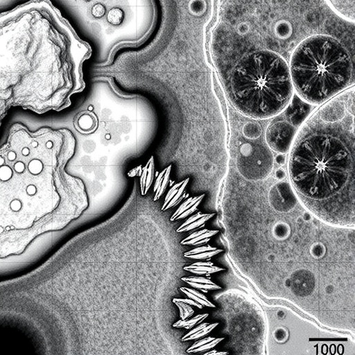
In a groundbreaking advancement poised to revolutionize the field of histology, researchers have unveiled a novel technique for mapping tissue fibers with micron-level precision that is completely independent of traditional sample preparation methods. This innovative approach, detailed in a recent publication in Nature Communications, signifies a transformative leap in the way scientists can analyze the microstructural organization of biological tissues, potentially unlocking new avenues in both medical diagnostics and fundamental biological research.
Histology, the microscopic study of tissue architecture, has long relied on complex and often labor-intensive sample preparation techniques. These conventional protocols, which frequently involve processes like sectioning, staining, and fixation, can introduce artifacts or alter the native structural properties of the fibers within tissues, obfuscating the true biological context. The new fiber mapping methodology circumvents these challenges entirely, offering a means to visualize and quantify fiber arrangements at an unprecedented resolution without the prerequisite of altering the tissue through preparation steps.
At the core of this breakthrough lies a fusion of advanced imaging modalities with sophisticated computational analysis. Through the integration of these modalities, the research team has been able to extract detailed fiber orientation and density information directly from histological samples in their native state. This capability not only preserves the integrity of the tissue but also ensures that the resulting data reflect authentic biological configurations, providing a vital tool for accurate assessments.
One critical aspect of this technique is its applicability across various sample types without the need for specific staining protocols, which historically limited the versatility of histological preparation. By rendering fiber visualization independent of staining, the method dramatically reduces the time and resources typically expended on sample preparation while simultaneously enhancing reproducibility. This could prove especially beneficial for large-scale studies or clinical settings where rapid processing times are paramount.
The researchers employed cutting-edge imaging hardware capable of capturing fine structural details at the micron scale. The resulting image data underwent rigorous computational processing, wherein algorithms meticulously analyzed fiber orientations, lengths, and intersections within the tissue matrix. This analytical procedure not only mapped fibers with remarkable accuracy but also allowed quantification of fiber properties that could be critical in understanding tissue mechanics and pathology.
Beyond its immediate histological applications, this innovation holds promise for elucidating complex biological phenomena. For instance, the precise mapping of collagen fibers and other extracellular matrix components could provide new insights into disease progression mechanisms such as fibrosis or cancer metastasis, where alterations in fiber architecture play pivotal roles. By enabling researchers to observe these microstructural changes unobscured by preparation-induced distortions, the technology may expedite breakthroughs in disease modeling and therapeutic development.
Moreover, this method could serve as a vital complement to existing imaging techniques, such as multiphoton microscopy or electron microscopy, by offering a rapid and less intrusive assessment tool. Its compatibility with a broad range of tissue types—including soft and hard tissues—enhances its applicability across domains such as neuroscience, oncology, and regenerative medicine. The ability to obtain highly detailed fiber maps from untouched samples accelerates hypothesis generation and validation in these fields.
One of the remarkable features of this approach is its non-destructive nature. Traditional histological processing often consumes or irreversibly modifies precious samples, limiting subsequent analyses. In contrast, this fiber mapping technique preserves the samples intact, allowing for longitudinal studies or parallel investigations using other methodologies on the same tissue piece. This preservation not only maximizes the utility of specimen repositories but also aligns with ethical imperatives to minimize sample consumption.
The implications extend into clinical diagnostics, where accurate tissue characterization is essential. The ability to map fibers with micron resolution without preparatory modifications could enhance biopsy evaluations, providing clinicians with richer datasets to inform diagnoses and treatment decisions. For diseases like cancer, where tissue microarchitecture can be prognostic, this technology offers a pathway to more nuanced and personalized assessments.
Beyond its scientific and clinical merits, the approach exemplifies technological ingenuity by harnessing algorithmic power to translate complex image data into biologically meaningful representations. This synergy between hardware and software represents a burgeoning trend in biomedical research, wherein computational techniques increasingly augment and sometimes redefine experimental possibilities. Such integration accelerates data interpretation and fosters the development of automated workflows to handle large volumes of imaging data.
As the scientific community digests this pioneering work, potential extensions of the method are already on the horizon. Researchers anticipate adapting the technique to three-dimensional fiber mapping, which would offer volumetric insights into tissue architecture and enable more comprehensive understanding of structural relationships within complex organs. These advancements could further revolutionize tissue analysis paradigms and open previously inaccessible dimensions of biological inquiry.
The accessibility and adaptability of this fiber mapping technology suggest it will soon become a staple in research laboratories worldwide. Its ability to be implemented independently of sample preparation intricacies democratizes high-resolution fiber analysis, enabling broader participation and fostering cross-disciplinary collaborations. As more groups adopt and refine the technique, its range of applications is likely to expand, driving innovation across multiple branches of life sciences.
In sum, this new method for micron-resolution fiber mapping heralds a new era in histological analysis marked by greater accuracy, efficiency, and biological fidelity. By liberating researchers from the constraints of traditional sample preparation, it empowers them to observe tissues as they exist naturally, thereby deepening our understanding of their fundamental structures and functions. The potential ripple effects across biomedical research and clinical practice promise to be profound, marking this innovation as a landmark achievement in the evolving landscape of tissue imaging.
The research indicates a compelling future where histological insights are limited not by preparation artifacts but by the boundaries of technological imagination. Through continued refinement and integration with other modalities, such fiber mapping techniques could redefine standards for tissue analysis and expand the frontiers of diagnostic precision and biological discovery. As such, this work not only addresses a longstanding challenge but also charts an inspiring trajectory for future innovations in microscopic tissue characterization.
Subject of Research:
Micron-scale mapping of tissue fiber architecture independent of traditional sample preparation methods.
Article Title:
Micron-resolution fiber mapping in histology independent of sample preparation.
Article References:
Georgiadis, M., auf der Heiden, F., Abbasi, H. et al. Micron-resolution fiber mapping in histology independent of sample preparation. Nat Commun 16, 9572 (2025). https://doi.org/10.1038/s41467-025-64896-9
Image Credits: AI Generated
DOI:
https://doi.org/10.1038/s41467-025-64896-9
Tags: advanced imaging modalities in histologycomputational analysis in biological researchfiber orientation and density measurementhistology without sample preparationinnovative histological methodsmicron-scale fiber mappingmicrostructural organization of tissuesnative tissue visualization techniquesnon-invasive tissue analysis methodsrevolutionary histology researchtissue fiber analysis techniquestransformative advances in medical diagnostics


