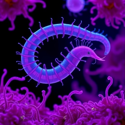
A groundbreaking study recently published in Science Advances on September 10 has reshaped our understanding of cellular environments within living multicellular organisms. A team of researchers from the University of California, Davis, has employed an innovative approach to track the movement of microscopic fluorescent particles inside the cells of Caenorhabditis elegans, a transparent nematode worm widely studied in developmental biology. This pioneering work reveals that the internal cytoplasmic environment of these worms is vastly more crowded and compartmentalized than previously understood from studies using single-celled yeast or cultured mammalian cells.
Traditional cellular biology has predominantly relied on in vitro systems—cultured mammalian cells or yeast—to investigate intracellular processes. However, the UC Davis team’s novel approach underscores a pivotal difference: cells within a living organism experience a highly constrained and spatially organized cytoplasm that cannot be fully replicated outside the physiological context. This finding challenges foundational assumptions about molecular diffusion and interaction made on the basis of cell culture studies and opens new avenues for understanding how cellular organization influences health and disease.
The researchers’ cutting-edge methodology centered on Genetically Encoded Multimeric Nanoparticles, or GEMs. These are engineered protein assemblies approximately 40 nanometers in diameter, roughly the size of a ribosome, designed to self-assemble and fluoresce inside living cells. By integrating DNA sequences encoding these GEMs into the C. elegans genome, scientists produced worms whose intestinal and epithelial cells endogenously assembled thousands of fluorescently tagged particles. Importantly, these genetically modified worms retained normal development and behavior, allowing for the monitoring of intracellular dynamics in a fully physiological context.
Using state-of-the-art time-lapse fluorescence microscopy capable of capturing movements at 50 frames per second, the team meticulously analyzed GEM mobility within the cytoplasm of living worms. Strikingly, they observed that particle movement was approximately 50-fold slower in worm cells compared to cultured mammalian or yeast cells. This dramatic reduction indicates a significantly denser and more structured intracellular milieu in multicellular organisms. Furthermore, the particles were not uniformly distributed but appeared to be confined within distinct subcellular compartments, defining a previously unappreciated level of spatial organization.
This profound level of compartmentalization prompted the investigators to delve into the molecular underpinnings maintaining cytoplasmic architecture. They identified a large scaffold protein, ANC-1—part of the KASH protein family—as a key player in structuring the cytoplasm. ANC-1 forms complexes that act as intracellular “boxes,” compartmentalizing the cytoplasm and enforcing spatial constraints on particle movement. Disruption of ANC-1 resulted in GEMs becoming more freely mobile within the cytoplasm, although overall crowding remained, indicating that ANC-1 governs spatial confinement rather than crowding density itself.
Complementing the ANC-1 scaffold system, the researchers highlighted the critical role of ribosomes in mediating cytoplasmic crowding. Ribosomes, known protein synthesis complexes, act like packing peanuts inside a box, filling the cytoplasm and reducing available free volume. When the team simultaneously disrupted both ANC-1 and ribosome production, GEM particles exhibited considerably increased mobility, suggesting that intracellular particle dynamics are governed by two complementary systems: the physical crowding by ribosomes and the spatial partitioning by ANC-1-mediated compartments.
These insights reveal a novel biophysical model for cytoplasmic organization in multicellular animals, where crowding and compartmentalization operate in concert to control molecular movement. By tightly regulating the mobility of macromolecular complexes, cells may fine-tune processes from signal transduction to metabolic flux, impacting facets of cell physiology previously inaccessible to direct observation. The findings carry profound implications for understanding drug delivery mechanisms and pathological states, including neurodegeneration and aging, where altered crowding and compartmentalization could disrupt cellular homeostasis.
The journey to implement GEMs into C. elegans was a formidable technical challenge, requiring years of iterative molecular engineering and optimization. The small size of the worm’s cells and the need for stable, endogenous expression of fluorescent nanoparticles posed significant hurdles overcome by the expertise of Starr, Luxton, Ding, and their colleagues. Their success in deploying GEMs in a living multicellular system represents a major technical milestone in cellular biophysics and highlights the immense potential of genetically encoded reporters for in vivo studies.
Looking ahead, the team plans to expand their use of GEM technology to examine neuron cells within C. elegans, given the critical relationship between cytoplasmic biophysical properties and neurodegenerative diseases. They are also preparing to introduce GEMs into more complex organisms such as zebrafish, broadening the scope to vertebrate systems where cellular architecture and crowding dynamics may differ further. This trajectory positions their research at the cutting edge of developmental genetics, biophysics, and disease biology.
As Luxton emphasizes, this approach transcends traditional cell culture paradigms by seeking to measure the physical properties of cells within their native organismal contexts. “The only way to truly understand how biophysical properties influence health is by directly observing living tissues,” he states. This paradigm shift has the potential to revolutionize how scientists investigate molecular interactions, intracellular transport, and the cellular environment’s role in disease progression and therapeutic response.
This study, enabled by advanced imaging at the UC Davis Molecular and Cellular Biology Light Microscopy Imaging Facility, was supported by the National Institutes of Health and the Paul G. Allen Frontiers Group. Its collaborative authorship includes multiple UC Davis researchers and collaborators from NYU Grossman School of Medicine, reflecting a multidisciplinary effort to push the boundaries of cell biology in living animals.
Ultimately, the discovery that multicellular organisms display a uniquely crowded and compartmentalized cytoplasm challenges long-held assumptions and opens a vibrant research frontier. It invites reevaluation of biochemical and physiological models developed in simplistic culture systems and underscores the rich complexity of living tissues. This work signals a dramatic shift in cellular biology, leveraging molecular engineering, biophysics, and advanced microscopy to uncover the hidden physical rules that govern life at the microscopic scale.
Subject of Research: Animals
Article Title: Giant KASH proteins and ribosomes establish distinct cytoplasmic biophysical properties in vivo
News Publication Date: 10-Sep-2025
Web References:
DOI link
Keywords: Cellular physiology, Cell metabolism, Cellular processes, Worms
Tags: Caenorhabditis elegans studycellular environmentscellular organization and healthcrowded cytoplasmic spacedevelopmental biology advancementsfluorescent particle trackinggenetically encoded multimeric nanoparticlesinnovative cellular biology methodsintracellular processesmolecular diffusion in cellsmulticellular organism researchphysiological context in research


