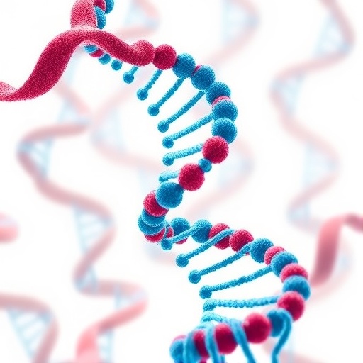
In the intricate realm of molecular biology, DNA is often visualized as the iconic double helix—a neatly twisted ladder representing the blueprint of life. Yet, inside every living cell, this molecule is far from tidy. DNA strands fold, twist, loop, and sometimes become tangled in complex knots, posing serious challenges to cellular function and genetic stability. Recent advances by an international team led by the University of Sheffield push the boundaries of our understanding of DNA topology, revealing a new frontier where the precise visualization of these tangled structures could illuminate fundamental biological processes and disease mechanisms.
At the heart of these discoveries lies a pioneering approach integrating atomic force microscopy (AFM), advanced imaging software, and artificial intelligence. Unlike traditional microscopes using light or electrons, AFM employs a nanoscale mechanical probe that physically “feels” the surface of molecules, generated topographical images with unprecedented nanometre (one-billionth of a meter) resolution. This technique enables researchers to capture the subtle twists and crossings in DNA strands, a feat previously unattainable with such accuracy and speed.
One of the remarkable breakthroughs is the ability to distinguish, at each point where DNA strands intersect, which strand is passing over and which passes under. This distinction may seem subtle, but it has profound implications for understanding the molecular mechanics inside cells. Each crossover influences how enzymes interact with DNA, especially topoisomerases, which play a critical role in resolving tangles and knots to maintain genomic integrity. Prior to this study, manually mapping these crossings was labor-intensive and prone to errors, significantly limiting the throughput of DNA topology research.
.adsslot_wEfD6mRWQp{width:728px !important;height:90px !important;}
@media(max-width:1199px){ .adsslot_wEfD6mRWQp{width:468px !important;height:60px !important;}
}
@media(max-width:767px){ .adsslot_wEfD6mRWQp{width:320px !important;height:50px !important;}
}
ADVERTISEMENT
The new automated method developed by the Sheffield-led group harnesses the power of AI to analyze AFM images swiftly and accurately. The software algorithms trace the convoluted paths of DNA molecules, quantifying their complexity and revealing structural nuances within seconds—tasks that would previously take scientists hours or even days. This high-resolution, automated analysis is instrumental in expanding our understanding of how DNA configuration affects biological processes like replication, transcription, and chromatin remodeling.
DNA topology is not merely a curiosity of molecular architecture but a crucial determinant of cellular health. When DNA becomes excessively knotted or tangled, vital processes can become obstructed, potentially triggering genomic instability. Such disruptions have been implicated in a wide array of diseases, including various cancers and neurodegenerative disorders. Therefore, the ability to scrutinize DNA at this level of detail may facilitate the identification of early pathological changes and aid the development of novel therapeutic strategies targeting DNA repair and maintenance pathways.
The collaborative research team also employed extensive molecular simulations to unravel how DNA interacts with surfaces used in AFM experiments, such as mica. These computational models generate thousands of molecular configurations, producing a rich dataset that trains AI systems to recognize and interpret complex DNA topologies in real experimental conditions. This synergy between simulation, AI, and microscopy exemplifies the power of interdisciplinary science in addressing biological complexity.
Professor Alice Pyne, a leading biophysicist at the University of Sheffield, underscores the significance of this advancement: “By determining the structure of individual, complex DNA assemblies with nanometre precision, we usher in a new era in molecular imaging. These tools enable us to investigate the intricate structures formed during critical cellular processes and understand their biological roles and consequences more deeply than ever before.”
Co-author Dr. Sean Colloms of the University of Glasgow adds that the ability to differentiate the “over” and “under” strands at each crossing allows for the discrimination between different knot configurations and their mirror images—information vital for studying how cellular machinery recognizes and processes DNA knots. Such knowledge is essential for understanding how enzymes like topoisomerases resolve DNA entanglements, ensuring genetic stability.
This research also highlights the distinctive advantages of AFM over other microscopy techniques. By physically probing the molecular surface, AFM circumvents limitations inherent to light or electron microscopy, such as the need for fluorescent labels or potential damage to delicate molecules. This makes AFM particularly suited for nanoscale biological investigations where preserving native structure is critical.
The international nature of this study, spanning institutions across the United Kingdom, Slovakia, and France, showcases the collective effort driving innovation in molecular genetics. The findings, detailed in the prestigious journal Nature Communications, represent a significant leap forward, equipping scientists with the means to decode the complex three-dimensional conformation of DNA in unprecedented detail.
By providing a clearer picture of DNA topology, this research not only enhances fundamental genetic knowledge but also has practical implications for medicine and biotechnology. Understanding how DNA knotting affects protein interactions could inform the design of targeted antibiotics and anti-cancer drugs, many of which function by modulating topoisomerase activity.
As DNA research evolves, this integration of cutting-edge microscopy, computational modeling, and AI sets a new standard for molecular analysis, promising to unlock mysteries previously beyond reach. The capacity to visualize and quantify DNA structures at the nanoscale heralds exciting opportunities for diagnosing disease, designing therapies, and comprehending the exquisite choreography of life’s most fundamental molecule.
Subject of Research: Cells
Article Title: Quantifying complexity in DNA structures with high resolution Atomic Force Microscopy
News Publication Date: 1-Jul-2025
Web References:
Nature Communications Article
DOI: 10.15131/shef.data.22633528.v2
Image Credits: University of Sheffield in Nature Communications
Keywords: DNA damage, DNA topology, Atomic Force Microscopy, molecular genetics, molecular imaging, topoisomerases, nanoscale analysis, AI in biology
Tags: advances in atomic force microscopyartificial intelligence in biological researchbreakthroughs in cellular function studieschallenges in genetic stabilityDNA strand interactionsDNA topology researchimaging techniques in geneticsinternational collaboration in genetic researchintricate DNA folding mechanismsmolecular biology innovationsunraveling genetic disease mechanismsvisualization of DNA structures


