In a groundbreaking study published in Pediatric Radiology, researchers have uncovered significant correlations between baseline terminal ileal imaging measurements and subsequent imaging outcomes in pediatric patients diagnosed with Crohn’s disease. This insight is particularly crucial as it sheds light on a previously underexplored area in the assessment of inflammatory bowel disease (IBD) in children. The findings from this cohort study emphasize the importance of precise imaging techniques for optimizing clinical outcomes and tailoring personalized treatment approaches.
Crohn’s disease, a chronic inflammatory condition affecting the gastrointestinal tract, poses unique challenges in pediatric populations. The disease can lead to severe complications, including strictures, fistulas, and growth delays. Early diagnosis and efficient monitoring of disease progression are essential for effective management. This study aims to enhance the diagnostic accuracy and predictability of treatment responses by utilizing advanced imaging modalities such as computed tomography (CT) and magnetic resonance imaging (MRI).
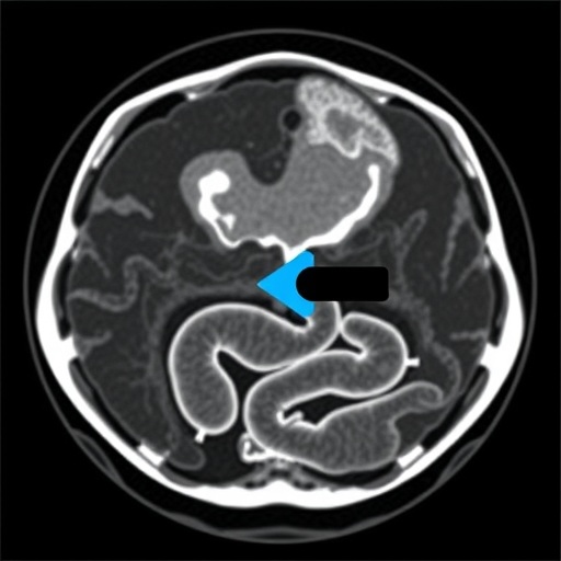
A focal point of the research lies in the evaluation of terminal ileal measurements obtained through these imaging techniques. The terminal ileum, the last part of the small intestine, is often a primary site of Crohn’s disease manifestation. By comparing baseline imaging data with clinical outcomes, the researchers have been able to establish a link that could pave the way for improved patient prognostication. This approach not only aids in understanding disease progression but may also influence treatment decisions.
.adsslot_F0OerqfzaA{width:728px !important;height:90px !important;}
@media(max-width:1199px){ .adsslot_F0OerqfzaA{width:468px !important;height:60px !important;}
}
@media(max-width:767px){ .adsslot_F0OerqfzaA{width:320px !important;height:50px !important;}
}
ADVERTISEMENT
The methodology implemented in this study is robust, involving a comprehensive cohort of pediatric patients diagnosed with Crohn’s disease. The researchers utilized data from various imaging studies conducted during the initial assessment of the patients. By analyzing measurements such as wall thickness, stricture formation, and inflammatory changes detected by CT and MRI, they worked to correlate these baseline metrics with clinical outcomes over time.
Another commendable aspect of the research is the integration of multidisciplinary expertise, combining the insights of radiologists, gastroenterologists, and pediatricians. This collaboration underscores the importance of a holistic approach to treating Crohn’s disease, highlighting how imaging can inform clinical practice. The study’s results are expected to enhance the communication between specialties, ensuring that patient care is both comprehensive and coordinated.
Furthermore, the implications of this study resonate beyond just individual patient outcomes. Establishing standardized imaging protocols for pediatric Crohn’s disease will likely improve the consistency of care across different healthcare settings. As clinicians adopt these findings, the emphasis on meticulous imaging may lead to earlier detection of complications and better long-term outcomes for children suffering from this distressing illness.
The use of advanced imaging technologies such as CT and MRI continues to evolve, offering enhanced visualizations that provide deeper insights into the pathophysiology of Crohn’s disease. By focusing on the terminal ileum, the study may inspire further research into how these imaging modalities can be utilized to monitor disease activity and treatment responses more effectively.
The potential for imaging findings to predict treatment outcomes paves the way for tailored therapeutic strategies. Physicians may soon have the ability to personalize treatment regimens based on specific imaging indicators, leading to more targeted therapies that minimize adverse effects and maximize efficacy. This patient-centered approach aligns with contemporary movements in medicine to prioritize individualized care.
As the medical community continues to grapple with the complexities of IBD, studies like this one not only illuminate critical aspects of disease management but also underscore the need for ongoing research. Further exploration into additional imaging parameters and their correlations with clinical variables may yield even more insightful revelations. The pursuit of scientific knowledge in this area is vital for informing future guidelines and practice standards.
Importantly, the study highlights that while imaging is a powerful tool for assessing Crohn’s disease, it is not without limitations. Factors such as patient compliance, variations in imaging protocols, and the subjective interpretation of results can influence outcomes. Acknowledging these challenges is crucial for researchers and clinicians as they work towards integrating these findings into clinical practice.
The role of educational programs to train healthcare professionals on the interpretation of imaging results in the context of Crohn’s disease cannot be overstated. As advanced imaging becomes central to pediatric gastroenterology, providing clinicians with the skills to interpret these findings accurately will be crucial in ensuring that patients receive optimal care.
Looking ahead, the integration of artificial intelligence (AI) and machine learning into imaging analysis has the potential to revolutionize this field further. By automating measurements and enhancing detection capabilities, AI could assist radiologists in identifying subtle changes that may be indicative of disease progression. This innovation could serve as a valuable adjunct to traditional imaging interpretations in pediatrics.
In conclusion, the findings from this cohort study represent a significant advancement in understanding the relationship between imaging measurements and outcomes in pediatric Crohn’s disease. As researchers continue to investigate the nuances of this condition, the contributions of imaging science will undoubtedly play a pivotal role in shaping the future landscape of IBD management. The insights gained from this research can empower clinicians to make informed decisions, ensuring that children with Crohn’s disease can lead healthier and more fulfilling lives.
As this study garners attention, it is imperative that the broader medical community embraces these findings and implements them into routine clinical practice for the benefit of pediatric patients everywhere. The promise of improved outcomes through enhanced imaging techniques lays a hopeful foundation for future advancements in the management of Crohn’s disease.
Subject of Research: Pediatric Crohn’s disease and imaging outcomes.
Article Title: Baseline terminal ileal CT and MRI measurements are associated with imaging outcomes in pediatric Crohn’s disease: a cohort study.
Article References:
Ta, A.D., Dillman, J.R., Ollberding, N.J. et al. Baseline terminal ileal CT and MRI measurements are associated with imaging outcomes in pediatric Crohn’s disease: a cohort study. Pediatr Radiol 55, 1642–1651 (2025). https://doi.org/10.1007/s00247-025-06302-6
Image Credits: AI Generated
DOI: July 2025
Keywords: Imaging, pediatric Crohn’s disease, CT, MRI, disease progression, treatment outcomes, individualized care, gastrointestinal health.
Tags: advanced imaging techniques in Crohn’s diseasechronic gastrointestinal conditions in childrencomplications of Crohn’s diseaseearly diagnosis of Crohn’s diseaseeffective management of inflammatory bowel diseaseimaging metrics in inflammatory bowel diseaseimaging outcomes in pediatric patientspediatric Crohn’s diseasepediatric radiology researchpersonalized treatment approaches for IBDterminal ileal measurementstreatment response predictability

