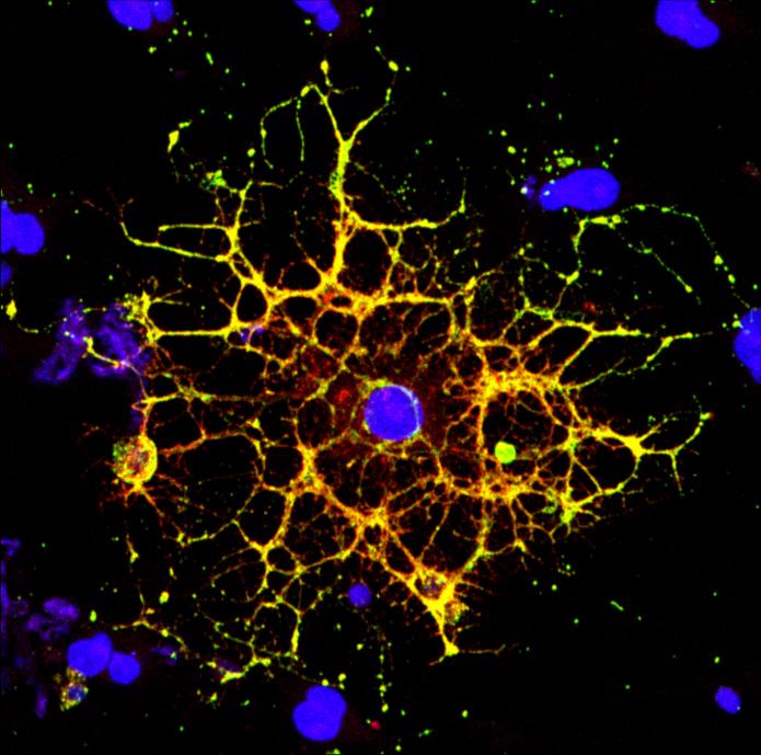
Researchers from Penn and CHOP detail the mechanism by which HIV infection blocks the maturation process of brain cells that produce myelin, a fatty substance that insulates neurons
It’s long been known that people living with HIV experience a loss of white matter in their brains. As opposed to “gray matter,” which is composed of the cell bodies of neurons, white matter is made up of a fatty substance called myelin that coats neurons, offering protection and helping them transmit signals quickly and efficiently. A reduction in white matter is associated with motor and cognitive impairment.
Earlier work by a team from the University of Pennsylvania and Children’s Hospital of Philadelphia (CHOP) found that antiretroviral therapy (ART)–the lifesaving suite of drugs that many people with HIV use daily–can reduce white matter, but it wasn’t clear how the virus itself contributed to this loss.
In a new study using both human and rodent cells, the team has hammered out a detailed mechanism, revealing how HIV prevents the myelin-making brain cells called oligodendrocytes from maturing, thus putting a wrench in white matter production. When the researchers applied a compound blocking this process, the cells were once again able to mature.
The work is published in the journal Glia.
“Even when people with HIV have their disease well-controlled by antiretrovirals, they still have the virus present in their bodies, so this study came out of our interest in understanding how HIV infection itself affects white matter,” says Kelly Jordan-Sciutto, a professor in Penn’s School of Dental Medicine and co-senior author on the study. “By understanding those mechanisms, we can take the next step to protect people with HIV infection from these impacts.”
“When people think about the brain, they think of neurons, but they often don’t think about white matter, as important as it is,” says Judith Grinspan, a research scientist at CHOP and the study’s other co-senior author. “But it’s clear that myelination is playing key roles in various stages of life: in infancy, in adolescence, and likely during learning in adulthood too. The more we find out about this biology, the more we can do to prevent white matter loss and the harms that can cause.”
Jordan-Sciutto and Grinspan have been collaborating for several years to elucidate how ART and HIV affect the brain, and specifically oligodendrocytes, a focus of Grinspan’s research. Their previous work on antiretrovirals had shown that commonly used drugs disrupted the function of oligodendrocytes, reducing myelin formation.
In the current study, they aimed to isolate the effect of HIV on this process. Led by Lindsay Roth, who recently earned her doctoral degree within the Biomedical Graduate Studies group at Penn and completed a postdoctoral fellowship working with Jordan-Sciutto and Grinspan, the investigation began by looking at human macrophages, one of the major cell types that HIV infects.
Scientists had hypothesized that HIV’s impact on the brain arose indirectly through the activity of these immune cells since the virus doesn’t infect neurons or oligodendrocytes. To learn more about how this might affect white matter specifically, the researchers took the fluid in which macrophages infected with HIV were growing and applied it to rat oligodendrocyte precursor cells, which mature into oligodendrocytes. While this treatment didn’t kill the precursor cells, it did block them from maturing into oligodendrocytes. Myelin production was subsequently also reduced.
“Immune cells that are infected with the virus secrete harmful substances, which normally target invading organisms, but can can also kill nearby cells, such as neurons, or stop them from differentiating,” Grinspan says. “So the next step was to figure out what was being secreted to cause this effect on the oligodendrocytes.”
The researchers had a clue to go on: Glutamate, a neurotransmitter, is known to have neurotoxic effects when it reaches high levels. “If you have too much glutamate, you’re in big trouble,” says Grinspan. Sure enough, when the researchers applied a compound that blunts glutamate levels to HIV-infected macrophages before the transfer of the growth medium to oligodendrocyte precursors, the cells were able to mature into oligodendrocytes. The result suggests that glutamate secreted by the infected macrophages was the culprit behind the precursor cells getting “stuck” in their immature form.
There was another mechanism, however, that the researchers suspected might be involved: the integrated stress response. This response integrates signals from four different signaling pathways, resulting in changes in gene expression that serve to protect the cell from stress or to prompt the cell to die, if the stress is overwhelming. Earlier findings from Jordan-Sciutto’s lab had found the integrated stress response was activated in other types of brain cells in patients who had cognitive impairment associated with HIV infection, so the team looked for its involvement in oligodendrocytes as well.
Indeed, they found evidence that the integrated stress response was activated in cultures of oligodendrocyte precursor cells.
Taking this information with what they had found out about glutamate, “Lindsay was able to tie these two things together,” Jordan-Sciutto says. She demonstrated that HIV-infected macrophages secreted glutamate, which activated the integrated stress response by turning on a pathway governed by an enzyme called PERK. “If you blocked glutamate, you prevented the activation of the integrated stress response,” Jordan-Sciutto says.
To take these findings further, and potentially test out new drug targets to address HIV-related cognitive impairments, the team hopes to use a well-characterized rat model of HIV infection.
“HIV is a human disease, so it’s a hard one to model,” says Grinspan. “We want to find out if this model recapitulates human disease more accurately than others we’ve used in the past.”
By tracking white matter in this animal model and comparing it to imaging studies done on patients with HIV, they hope to get at a better understanding of what factors shape white matter loss. They’re particularly interested in looking at a cohort of adolescents being treated at CHOP, as teens are a group in whom HIV infection rates are climbing.
Ultimately, the researchers want to discern the effects of the virus from the drugs used to treat it in order to better evaluate the risks of each.
“When we put people on ART, especially kids or adolescents, it’s important to understand the implications of doing that,” says Jordan-Sciutto. “Antiretrovirals may prevent the establishment of a viral reservoir in the central nervous system, which would be wonderful, but we also know that the drugs can cause harm, particularly to white matter.
“And then of course we can’t forget the 37 million HIV-infected individuals who live outside the United States and may not have access to antiretrovrials like the patients here,” she says. “We want to know how we can help them too.”
###
Kelly Jordan-Sciutto is vice chair and professor in the University of Pennsylvania School of Dental Medicine’s Department of Basic & Translational Sciences and is director of Biomedical Graduate Studies.
Judith Grinspan is research scientist at the Children’s Hospital of Philadelphia and research professor of neurology at the the Perelman School of Medicine at the University of Pennsylvania.
Lindsay Roth, who recently earned her doctoral degree from the Biomedical Graduate Group at the University of Pennsylvania, was first author on the paper.
Roth, Grinspan, and Jordan-Sciutto’s coauthor was Çagla Akay-Espinoza, from Penn’s School of Dental Medicine.
The study was supported by the National Institutes of Health (grants MH098742, MH118121, and MH109382) and the Cellular Neuroscience Core of the Institutional Intellectual and Developmental Disabilities Research Core of the Children’s Hospital of Philadelphia (grants HD26979 and GM008076).
Media Contact
Katherine Unger Baillie
[email protected]
Original Source
http://penntoday.
Related Journal Article
http://dx.


