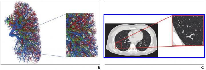
Computational patient models and human phantom with coronavirus abnormalities via multidiagnostic confirmation of SARS-CoV-2 infection yield ‘realistic’ texture and shape

Credit: American Roentgen Ray Society (ARRS), American Journal of Roentgenology (AJR)
Leesburg, VA, August 25, 2020–An open-access article in ARRS’ American Journal of Roentgenology (AJR) established a foundation for the use of virtual imaging trials in effective assessment and optimization of CT and radiography acquisitions and analysis tools to help manage the coronavirus disease (COVID-19) pandemic.
Virtual imaging trials have two main components–representative models of targeted subjects and realistic models of imaging scanners–and the authors of this AJR article developed the first computational models of patients with COVID-19, while showing, as proof of principle, how they can be combined with imaging simulators for COVID-19 imaging studies.
“For the body habitus of the models,” lead author Ehsan Abadi explained, “we used the 4D extended cardiac-torso (XCAT) model that was developed at Duke University.”
Abadi and his Duke colleagues then segmented the morphologic features of COVID-19 abnormalities from 20 CT images of patients with multidiagnostic confirmation of SARS-CoV-2 infection and incorporated them into XCAT models.
“Within a given disease area, the texture and material of the lung parenchyma in the XCAT were modified to match the properties observed in the clinical images,” Abadi et al. continued.
Using a specific CT scanner (Definition Flash, Siemens Healthineers) and validated radiography simulator (DukeSim) to help illustrate utility, the team virtually imaged three developed COVID-19 computational phantoms.
“Subjectively,” the authors concluded, “the simulated abnormalities were realistic in terms of shape and texture,” adding their preliminary results showed that the contrast-to-noise ratios in the abnormal regions were 1.6, 3.0, and 3.6 for 5-, 25-, and 50-mAs images, respectively.
###
Founded in 1900, the American Roentgen Ray Society (ARRS) is the first and oldest radiological society in North America, dedicated to the advancement of medicine through the profession of radiology and its allied sciences. An international forum for progress in medical imaging since the discovery of the x-ray, ARRS maintains its mission of improving health through a community committed to advancing knowledge and skills with an annual scientific meeting, monthly publication of the peer-reviewed American Journal of Roentgenology (AJR), quarterly issues of InPractice magazine, AJR Live Webinars and Podcasts, topical symposia, print and online educational materials, as well as awarding scholarships via The Roentgen Fund®.
Media Contact
Logan K. Young
[email protected]
Original Source
https:/
Related Journal Article
http://dx.


