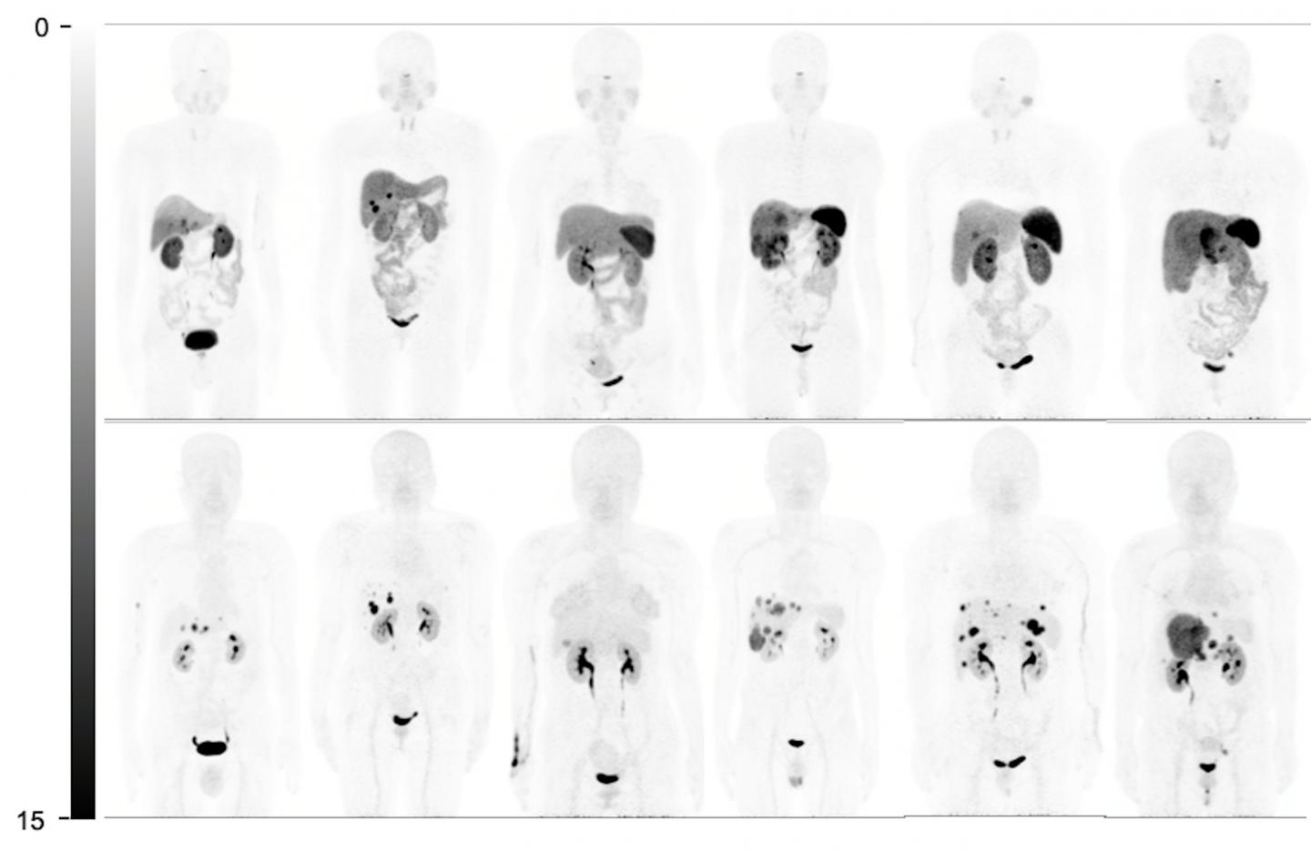
Imaging excellence shown in patients with liver-dominant disease

Credit: Images created by Wenjia Zhu and Li Huo, et al., Peking Union Medical College Hospital, Beijing, China.
Reston, VA–For neuroendocrine cancer patients with liver metastases, a new radiopharmaceutical, 68Ga-DOTA-JR11, has shown excellent imaging performance in tumor detection, staging and restaging, providing important information to guide treatment. In a head-to-head comparison of two somatostatin receptor (SSTR) imaging agents, 68Ga-DOTA-JR11 PET/CT performed better than 68Ga-DOTATATE PET/CT in detecting liver metastases, with a better tumor-to-background ratio, according to research published in the June issue of The Journal of Nuclear Medicine.
SSTRs–the key target for imaging and peptide receptor radionuclide therapy in patients with neuroendocrine tumors–typically are imaged using 68Ga-labeled peptides, which are agonists that bind to SSTRs to elicit a response. However, newly developed peptide antagonists, which recognize and then block SSTRs, have shown more favorable pharmacokinetics, better image contrast, higher tumor uptake, and better residence time in recent studies.
“With antagonists, we now have an alternative to agonists,” stated Wenjia Zhu, MD, nuclear medicine physician, at Peking Union Medical College Hospital in Bejing, China. “However, there is still not much evidence about the performance of PET/CT imaging with SSTR antagonists. Hence, we designed this prospective study to compare 68Ga-DOTATATE and 68Ga-DOTA-JR11 PET/CT in patients with metastatic, well-differentiated neuroendocrine tumors.”
The study included 31 patients and took place on two consecutive days. Each patient received an intravenous injection of 68Ga-DOTATATE on the first day and 68Ga-DOTA-JR11 on the second day. Whole-body time-of-flight PET/CT scans were performed 40-60 minutes after each injection on the same scanner. Upon completion, physiologic normal-organ uptake, lesion numbers, and lesion uptake were compared between 68Ga-DOTATATE and 68Ga-DOTA-JR11 PET/CT images.
The physiologic normal-organ uptake of the spleen, renal cortex, adrenal glands, pituitary glands, stomach wall, normal liver parenchyma, small intestine, pancreas, and bone marrow was significantly lower on 68Ga-DOTA-JR11 PET/CT than on 68Ga-DOTATATE PET/CT. 68Ga-DOTA-JR11 was found to detect significantly more liver lesions than 68Ga-DOTATATE; however 68Ga-DOTATATE detected more bone lesions than 68Ga-DOTA-JR11. While the radiopharmaceuticals showed similar lesion uptake for primary tumors and lymph node metastases on both patient-based and lesion-based comparisons, the target-to-background ratio of liver lesions was significantly higher on 68Ga-DOTA-JR11.
“What we’ve learned from this study is that peptides matter,” noted Zhu. “For patients with different metastatic patterns, different peptides (DOTA-JR11 versus DOTATATE) should be used. In liver-dominant disease, 68Ga-DOTA-JR11 may be a better choice in tumor staging and restaging compared to 68Ga-DOTATATE. It may also change the treatment strategy, especially when partial resection or local therapy for liver metastasis is considered. In bone-dominant disease, we should probably stick to agonists, as 68Ga-DOTA-JR11 may underestimate tumor burden. We expect more extensive theranostic application of antagonists in the near future.”
###
The authors of “Head-to-Head Comparison of 68Ga-DOTA-JR11 and 68Ga-DOTATATE PET/CT in Patients with Metastatic, Well-Differentiated Neuroendocrine Tumors: A Prospective Study” include Wenjia Zhu, Xuezhu Wang and Li Huo, Beijing Key Laboratory of Molecular Targeted Diagnosis and Therapy in Nuclear Medicine, Department of Nuclear Medicine, Peking Union Medical College Hospital, Chinese Academy of Medical Sciences and Peking Union Medical College, Beijing, China; Yuejuan Cheng and Chunmei Bai, Department of Oncology, Peking Union Medical College Hospital, Beijing, China; Shaobo Yao, Department of PET/CT Diagnostics, General Hospital, Tianjin Medical University Tianjin, China; Hong Zhao, Department of Hepatobiliary Surgery, National Cancer Center/National Clinical Research Center for Cancer/Cancer Hospital, Chinese Academy of Medical Sciences and Peking Union Medical College, Beijing, China; and Ru Jia and Jianming Xu, Department of Gastrointestinal Oncology, Fifth Medical Center, General Hospital of PLA, Beijing, China.
This study was made available online in November 2019 ahead of final publication in print in June 2020.
Please visit the SNMMI Media Center for more information about molecular imaging and precision imaging. To schedule an interview with the researchers, please contact Rebecca Maxey at (703) 652-6772 or [email protected].
About the Society of Nuclear Medicine and Molecular Imaging
The Journal of Nuclear Medicine (JNM) is the world’s leading nuclear medicine, molecular imaging and theranostics journal, accessed close to 10 million times each year by practitioners around the globe, providing them with the information they need to advance this rapidly expanding field. Current and past issues of The Journal of Nuclear Medicine can be found online at http://jnm.
JNM is published by the Society of Nuclear Medicine and Molecular Imaging (SNMMI), an international scientific and medical organization dedicated to advancing nuclear medicine and molecular imaging–precision medicine that allows diagnosis and treatment to be tailored to individual patients in order to achieve the best possible outcomes. For more information, visit http://www.
Media Contact
Rebecca Maxey
[email protected]
Original Source
https:/
Related Journal Article
http://dx.


