Credit: Images courtesy of the GSK Center for Optical Molecular Imaging
Researchers at the GSK Center for Optical Molecular Imaging have developed a new microscope that looks at the different parameters that change during wound healing. They hope to use this technique to understand how skin disorders, such as foot ulcers in diabetic patients and psoriasis, can be treated.
“Nobody really understands how topical drugs affect the skin because they can’t see below the skin,” said Marina Marjanovic, an associate professor of bioengineering and associate director of the center, which is located at the Beckman Institute for Advanced Science and Technology at the University of Illinois at Urbana-Champaign.
“We need this technique to understand whether the available treatments are curing the underlying condition or just the symptoms,” said Marjanovic, who also is a member of Beckman’s Biophotonics Imaging Lab.
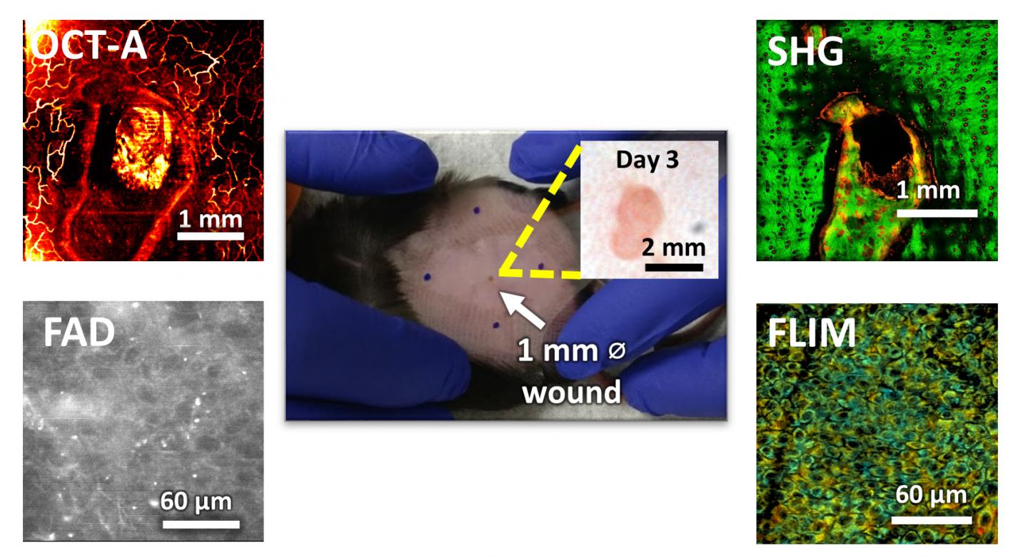
The new microscope can look at different aspects of wound healing simultaneously. The researchers used it to get images of the wound, track collagen that helps in wound healing, and visualize the blood vessel distribution around the wound. Additionally, they also have measured various chemicals in the tissue that indicate how much inflammation is occurring.
The paper “Non-invasive monitoring of pharmacodynamics during the skin wound healing process using multimodal optical microscopy” was published in BMJ Open Diabetes Research & Care.
“This is a continuation of a previous study we did where we made a wound on the ears of mice,” said Aneesh Alex, a visiting scholar at the Beckman Institute and a scientist at GlaxoSmithKline. “In this study, we added more visualization capabilities and studied the wound healing process on the backs of the mice, which is a more accurate model.”
The researchers studied the healing process for a month. They looked at four groups of mice: mice with untreated wounds, mice which had placebo treatments, and mice with two different concentrations of the treatment drug.
“One of the main limitations of optical imaging techniques is its shallow penetration depth in biological tissues,” said Eric Chaney, a research scientist in the Biophotonics Imaging Lab and at the center. The limitation is due to the scattering of light, which makes it difficult for the researchers to look at deeper tissue structures.
“The major advantages of this technique are that we do not use any labels in our imaging and it is completely noninvasive,” Marjanovic said. “We have also done follow-up studies in humans and we have been able to look at the changes in the skin of healthy volunteers and patients with skin conditions.”
The researchers hope to further refine the technique so it can be used for routine studies of skin disorders and their treatments.
“Our Center for Optical Molecular Imaging at the Beckman Institute has been a unique and productive academic-industry partnership with GlaxoSmithKline,” said Dr. Stephen Boppart, Abel Bliss Professor of Engineering and a professor of electrical and computer engineering and of bioengineering, who also is a medical doctor. “By combining our state-of-the-art optical imaging technologies, we have the ability to not only visualize and understand the molecular interactions that occur, but also how these processes may affect drug action and efficacy.”
The work was done in collaboration with GlaxoSmithKline. The data analysis was done with the help of Salma Musaad, a research biostatistician at the Interdisciplinary Health Sciences Institute.
The paper “Non-invasive monitoring of pharmacodynamics during the skin wound healing process using multimodal optical microscopy” can be found at http://dx.
Media Contact
Doris Dahl
[email protected]
Original Source
https:/
Related Journal Article
http://dx.
