Latest breakthrough by Professor Sylvain Martel’s team at Polytechnique Montréal opens up new horizons for endovascular surgery

Credit: Massouh bioMEDia for the Polytechnique Montréal Nanorobotics Laboratory
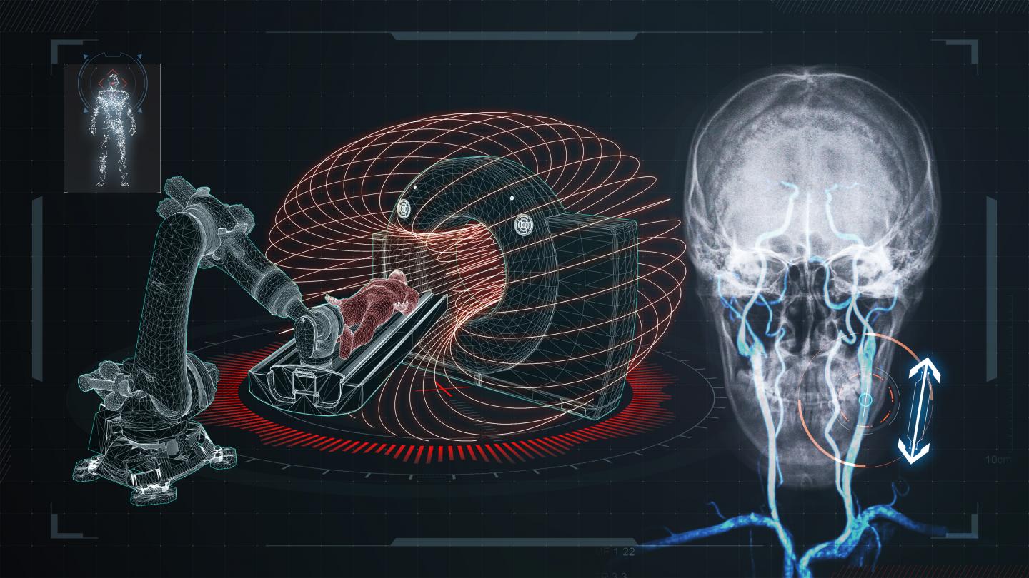
A team led by Professor Sylvain Martel at the Polytechnique Montréal Nanorobotics Laboratory has developed a novel approach to tackling one of the biggest challenges of endovascular surgery: how to reach the most difficult-to-access physiological locations. Their solution is a robotic platform that uses the fringe field generated by the superconducting magnet of a clinical magnetic resonance imaging (MRI) scanner to guide medical instruments through deeper and more complex vascular structures. The approach has been successfully demonstrated in-vivo, and is the subject of an article just published in Science Robotics.
When a researcher “thinks outside the box”–literally
Imagine having to push a wire as thin as a human hair deeper and deeper inside a very long, very narrow tube full of twists and turns. The wire’s lack of rigidity, along with the friction forces exerted on the walls of the tube, will eventually render the manoeuvre impossible, with the wire ending up folded on itself and stuck in a turn of the tube. This is exactly the challenge facing surgeons who seek to perform minimally invasive procedures in ever-deeper parts of the human body by steering a guidewire or other instrumentation (such as a catheter) through narrow, tortuous networks of blood vessels.
It is possible, however, to harness a directional pulling force to complement the pushing force, countering the friction forces inside the blood vessel and moving the instrument much farther. The tip of the device is magnetized, and pulled along inside the vessels by the attraction force of another magnet. Only a powerful superconducting magnet outside the patient’s body can provide the extra attraction needed to steer the magnetized device as far as possible. There is one piece of modern hospital equipment that can play that role: an MRI scanner, which has a superconducting magnet that generates a field tens of thousands of times stronger than that of the Earth.
The magnetic field inside the tunnel of an MRI scanner, however, is uniform; this is key to how patient imaging is performed. That uniformity poses a problem: to pull the tip of the instrument through the labyrinthine vascular structures, the guiding magnetic field must be modulated to the greatest possible amplitude and then be decreased as quickly as possible.
Pondering that problem, Professor Martel had the idea of using not the main magnetic field present inside the MRI machine tunnel, but the so-called fringe field outside the machine. “Manufacturers of MRI scanners will normally reduce the fringe field to the minimum,” he explains. “The result is a very-high-amplitude field that decays very rapidly. For us, that fringe field represents an excellent solution that is far superior to the best existing magnetic guidance approaches, and it is in a peripheral space conducive to human-scale interventions. To the best of our knowledge, this is the first time that an MRI fringe field has been used for a medical application,” he adds.
Move the patient rather than the field
To steer an instrument deep within blood vessels, not only is a strong attraction force required, but that force must be oriented to pull the magnetic tip of the instrument in various directions inside the vessels. Because of the MRI scanner’s size and weight, it’s impossible to move it to change the direction of the magnetic field. To get around that issue, the patient is moved in the vicinity of the MRI machine instead. The platform developed by Professor Martel’s team uses a robotic table positioned within the fringe field next to the scanner.
The table, designed by Arash Azizi–the lead author of the article and a biomedical engineering PhD candidate whose thesis advisor is Professor Martel–can move on all axes to position and orient the patient according to the direction in which the instrument must be guided through their body. The table automatically changes direction and orientation to position the patient optimally for the successive stages of the instrument’s journey thanks to a system that maps the directional forces of the MRI scanner’s magnetic field–a technique that Professor Martel has dubbed Fringe Field Navigation (FFN).
An in-vivo study of FFN with X-ray mapping demonstrated the capacity of the system for efficient and minimally invasive steering of extremely small-diameter instruments deep within complex vascular structures that were hitherto inaccessible using known methods.
Robots to the rescue of surgeons
This robotic solution, which greatly outperforms manual procedures as well as existing magnetic field-based platforms, enables endovascular interventional procedures in very deep, and therefore currently inaccessible, regions of the human body.
The method promises to broaden possibilities for application of various medical procedures including diagnosis, imaging and local treatments. Among other things, it could serve to assist surgeons in procedures requiring the least invasive methods possible, including treatment of brain damage such as an aneurysm or a stroke.
###
This research work received support from the Canada Research Chairs Program.
About Polytechnique Montréal
Founded in 1873, Polytechnique Montréal, a technological university, is one of Canada’s largest engineering education and research institutions, and ranks first in Québec in terms of the scope of its engineering research activities. It is located on the Université de Montreal campus, the largest French-language university campus in the Americas. With over 50,000 graduates to this day, Polytechnique Montréal has educated 22% of the Ordre des ingénieurs du Québec’s current membership. Polytechnique offers more than 120 programs, taught by 280 professors and welcomes 9,000 students yearly, and has an annual operating budget of $260 million, including a research budget of $100 million.
DOI: 10.1126/scirobotics.aax7342
Paper: https:/
Media Contact
Annie Touchette
[email protected]
514-231-8133
Original Source
https:/
Related Journal Article
http://dx.


