In a remarkable stride toward revolutionizing liver disease diagnostics, researchers have unveiled a pioneering 3D multiparametric ultrasound imaging technique that dramatically enhances the detection and characterization of steatotic liver disease. This advancement, chronicled in a 2025 study led by Lee, D., Heo, J., Mun, H., and colleagues, represents a significant leap from traditional imaging modalities, leveraging the power of three-dimensional visualization combined with multiparametric data to unravel the complexities of fat accumulation in liver tissues.
Steatotic liver disease, encompassing a spectrum from simple fatty liver to non-alcoholic steatohepatitis (NASH), poses an escalating global health challenge due to its asymptomatic progression and potential to culminate in cirrhosis or hepatocellular carcinoma. Conventional diagnostic tools, primarily biopsy and two-dimensional ultrasound, face limitations either due to invasiveness or restricted spatial resolution. The cutting-edge 3D multiparametric ultrasound approach addresses these shortcomings by providing a comprehensive, non-invasive view of liver pathology with enhanced diagnostic accuracy.
At the heart of this innovation lies the fusion of three-dimensional volumetric imaging with multiple ultrasound-based parameters, including tissue stiffness, perfusion, and fat content assessment. This integrative methodology empowers clinicians and researchers to assess the liver’s structural and functional status simultaneously, thereby achieving a nuanced understanding of disease progression at an early stage. The technology’s multi-channel capability captures detailed acoustic signals, converting them into vivid 3D renderings that map the heterogeneity of hepatic tissue affected by steatosis.
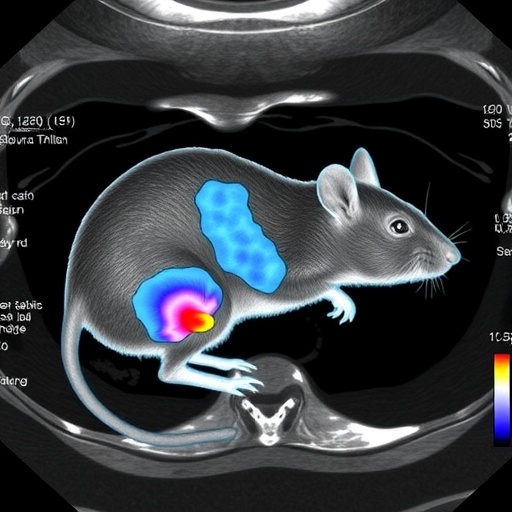
The experimental validation of this technique was meticulously conducted using male rat models, carefully selected to replicate human steatotic liver conditions. These in vivo studies revealed the system’s capacity to identify subtle changes in liver morphology and function, inaccessible through standard imaging. Notably, the multiparametric ultrasound modality detected variations in echogenicity and elasticity correlating directly with the severity of lipid infiltration and inflammation within hepatic tissues.
One of the remarkable technical achievements of this study is the optimization of ultrasound probe design and signal processing algorithms, which together enable enhanced penetration depth and spatial resolution. These advances mitigate common issues such as acoustic shadowing and speckle noise, prevalent challenges in ultrasound imaging of obese or fatty liver tissues. The 3D reconstruction algorithms synthesize the multiparametric data sets into coherent volumetric images, facilitating both qualitative assessment and quantitative analysis with unprecedented precision.
The implications of this technology stretch beyond mere diagnostics. By accurately mapping steatotic regions and providing real-time feedback on tissue characteristics, this ultrasound platform paves the way for personalized therapeutic interventions. Clinicians can now monitor treatment efficacy dynamically, adjusting regimens based on direct imaging evidence of liver tissue response. This could fundamentally transform patient management, reducing reliance on invasive liver biopsy and enhancing long-term outcomes.
Moreover, the non-invasive nature and relative affordability of ultrasound compared to magnetic resonance imaging (MRI) or computed tomography (CT) make this innovation particularly appealing for widespread clinical adoption. Its ability to provide rapid, bedside assessments aligns perfectly with the growing push for point-of-care diagnostics in hepatology, especially in resource-limited settings where advanced imaging infrastructure is scarce.
The data acquisition protocol, meticulously refined during this study, ensures reproducibility and consistency across scans, a critical factor for longitudinal patient monitoring. By integrating sophisticated motion correction algorithms, the system compensates for respiratory and cardiac-induced liver movements, thus preserving image fidelity and reducing artifacts commonly encountered in ultrasound imaging.
Importantly, this multiparametric ultrasound imaging modality extends its utility to fundamental research contexts, offering new windows into the pathophysiology of steatotic liver disease. Researchers can study dynamic tissue changes and microvascular alterations associated with fat accumulation and inflammatory processes in vivo, accelerating the discovery of novel biomarkers and therapeutic targets.
The interdisciplinary collaboration underpinning this breakthrough involved experts in biomedical engineering, hepatology, and computational imaging, reflecting a paradigm where technological innovation converges with clinical necessity. The combination of engineering prowess and medical insight was crucial to overcoming the complex acoustic challenges posed by the liver’s heterogeneous, fatty tissue environment.
Through rigorous validation against histopathological findings, the imaging parameters extracted from the multiparametric ultrasound strongly correlated with established markers of hepatic steatosis and fibrosis. This correlation underscores the system’s potential as a surrogate for biopsy, enabling safer serial monitoring of disease progression or regression in response to lifestyle modifications or pharmacologic treatment.
Future iterations of this technology aim at integrating artificial intelligence-driven image analysis, automating segmentation, and classification of affected liver zones. Such enhancements would further reduce operator dependency and improve diagnostic throughput, facilitating scalable deployment in clinical settings worldwide.
The study’s findings represent a seminal step toward democratizing liver disease diagnostics, bridging the gap between advanced imaging science and practical, accessible healthcare solutions. As liver disease prevalence continues to rise globally due to lifestyle factors and metabolic syndromes, innovations like this 3D multiparametric ultrasound imaging technique could not be more timely.
In summation, the integration of three-dimensional imaging with multiparametric ultrasound parameters has inaugurated a new era in hepatology, promising safer, more accurate, and comprehensive assessment of steatotic liver disease. The potential to transform diagnostic pathways, personalize treatment, and deepen our understanding of liver pathologies positions this technology at the frontier of medical imaging innovation.
Lee et al.’s contribution to the field poignantly illustrates how sophisticated engineering solutions can translate into tangible clinical benefits, heralding a future where liver disease is detected earlier, managed more effectively, and ultimately, outcomes are vastly improved for millions afflicted worldwide.
Subject of Research:
3D Multiparametric ultrasound imaging for the evaluation of steatotic liver disease.
Article Title:
3D multiparametric ultrasound imaging of steatotic liver disease in a study with male rats.
Article References:
Lee, D., Heo, J., Mun, H. et al. 3D multiparametric ultrasound imaging of steatotic liver disease in a study with male rats. Nat Commun 16, 10226 (2025). https://doi.org/10.1038/s41467-025-65046-x
Image Credits:
AI Generated
DOI:
https://doi.org/10.1038/s41467-025-65046-x
Tags: 3D multiparametric ultrasound imagingadvanced liver imaging techniquesfatty liver disease detectionhepatocellular carcinoma risk factorsinnovative medical imaging technologiesliver disease progression assessmentliver pathology evaluation methodsnon-alcoholic steatohepatitis (NASH) researchnon-invasive liver diagnosticssteatotic liver disease characterizationultrasound tissue stiffness measurementvolumetric imaging in liver diagnostics


