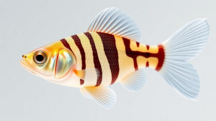
In a quiet laboratory, a tiny translucent fish embryo beats its heart for the first time. What looks like a simple biological milestone is, at the molecular level, an extraordinarily intricate performance choreographed by epigenetic machinery. A new review published in Epigenetics Communications by Pratiksha S. Kavade, Saili S. Parab, Vincenza Capone, Daniela Carannante, Concetta Ambrosino, Lucia Altucci and Vincenzo Carafa brings together decades of evidence showing that Polycomb group proteins, the cell’s master silencers, are indispensable for building both the zebrafish heart and its pectoral fins. These findings matter far beyond the aquarium, because the same genetic circuits that pattern a fish’s fins also shape human limbs, and the same epigenetic switches that form a two-chambered fish heart govern the four-chambered organ beating in our own chests.
Polycomb group proteins first announced themselves in fruit flies, where researchers screening for mutants noticed animals with an extra sex comb, a structure normally restricted to the front legs of males. At least sixteen of these genes were eventually identified as repressors of the homeotic, or Hox, genes, the ancient genetic address system that tells every cell where it sits along the body axis. The discovery revealed a profound biological principle: development depends not only on turning genes on, but on keeping the wrong genes firmly off. That principle has been conserved across hundreds of millions of years of evolution, and in vertebrates Polycomb proteins now appear central to cellular differentiation, proliferation, tissue integrity, homeostasis and stem cell renewal.
The molecular machinery is elegantly modular. Polycomb proteins assemble into two major multi-protein complexes, Polycomb Repressive Complex 1 and Polycomb Repressive Complex 2, each built around a catalytic core plus a variable set of accessory subunits. The catalytic heart of PRC1 is a heterodimer of RING1A or RING1B with one of six PCGF family members, and it stamps histone H2A with a single ubiquitin molecule at lysine 119, a mark abbreviated H2AK119ub. PRC2, whose catalytic engine is the methyltransferase EZH1 or EZH2 working alongside EED, SUZ12 and RBBP4/7, deposits di- and trimethyl marks on lysine 27 of histone H3, known as H3K27me2 and H3K27me3. The two complexes are deeply interdependent. The H3K27me3 mark laid down by PRC2 is recognised by the chromobox subunit of canonical PRC1, which is thereby recruited to the same target sites to spread H2AK119ub and lock genes into a repressed state. Variant PRC1 can operate in the opposite direction, ubiquitinating H2A first and pulling PRC2 in behind it, creating a self-reinforcing loop of silencing.
Studying these complexes in mammals is brutally difficult because embryos lacking Polycomb function typically die around gastrulation, long before organs form. This is precisely where the zebrafish, Danio rerio, earns its reputation as one of developmental biology’s most powerful model organisms. Introduced as a genetic system by George Streisinger in the 1970s, zebrafish combine optical clarity, rapid development, sexual maturity within twelve weeks of fertilisation, ex utero embryonic growth that allows manipulation from the single-cell stage, and clutches of hundreds of offspring per mating. Crucially, maternal gene products sustain the embryo up to roughly the 1000-cell stage, far longer than in mice, giving researchers an extended window to probe early gene regulation. Zebrafish embryos also survive early development even with severe cardiovascular defects, because they obtain oxygen by passive diffusion rather than relying solely on blood circulation, something no mammal can do.
The fish genome adds another layer of opportunity. Around 320 million years ago, the teleost lineage underwent a whole-genome duplication event, generating pairs of duplicate genes called ohnologs, of which roughly 15 to 20 percent were retained after rediploidization. In the zebrafish genome, thirteen Polycomb subunits exist as duplicated pairs, although some, including RING1A, PCGF2, PCGF3, RBBP7 and the accessory subunit HDAC2, were lost during rediploidization. Despite these losses, the diversity and complexity of Polycomb complexes remain intact, and researchers can now dissect their roles using an arsenal of techniques ranging from classical ENU mutagenesis and morpholino knockdowns to zinc-finger nucleases, TALENs and CRISPR/Cas9 gene editing.
When it comes to the heart, the review paints a picture of two complexes with distinct but complementary timing. Embryos lacking either PRC1 or PRC2 develop the striking ‘heart-strings’ phenotype: after 48 hours post-fertilisation, the heart fails to loop and instead remains a string-like tube rather than forming its two chambers, the atrium and ventricle. Loss of rnf2, the sole zebrafish homolog of PRC1’s enzymatic subunit, produces pericardial edema and this stringy heart. Yet early cardiac markers such as nkx2.5, tbx5 and hoxb5b appear at normal levels in the lateral plate mesoderm of rnf2 mutants, indicating that the heart field is specified correctly and that PRC1 acts later in the programme. RNA sequencing of individual mutant hearts by Chrispijn and colleagues showed that T-box transcription factors tbx2a, tbx2b, tbx3a and tbx5 become upregulated over time, while structural genes such as vmhc, myl7, myh6 and nppa are downregulated, pointing to a role for PRC1 in the structural maturation of the organ.
The finer details are remarkable. In homozygous rnf2(f5) mutants, genes involved in smooth muscle and skeletal muscle development, which normally participate in transforming the atrioventricular canal between the two chambers, become dysregulated. Transmission electron microscopy reveals misaligned cardiac sarcomeres, with closely packed myofibrils and abnormally spaced I-bands and Z-discs. Calcium imaging with the fluorescent dye fluo-4 AM shows a weakened calcium signal, and the result is a heart that contracts feebly. The expression of atrioventricular canal markers such as alcama, vcana and bmp4 becomes expanded and diffuse rather than tightly constricted. Together these data establish that PRC1 is essential for cardiac sarcomere assembly, the maintenance of contraction, and the proper constriction of the atrioventricular canal and its valves, largely by repressing inappropriate non-cardiac sarcomere genes. A second non-canonical PRC1 component, mga, adds another twist: morpholino knockdown of mgaa blocks heart tube looping, and the defect can be rescued by co-reducing gata4, whose transcript levels rise two- to four-fold in the mutant, showing that variant PRC1 fine-tunes this key regulator.
PRC2, by contrast, acts earlier. Maternal-zygotic ezh2 mutants, in which the methyltransferase is eliminated from both maternal and zygotic sources, show reduced numbers of nkx2.5-expressing cardiac progenitors, partially reduced chamber markers, and cardiomyocytes that wander away from the cardiac tube, leaving a small, tubular heart that never loops. The atrioventricular canal fails to form properly, possibly because of ectopic expression of has2, a gene that promotes cardiac cell migration. Chromatin immunoprecipitation sequencing shows that both ezh2 and the H3K27me3 mark are absent from heart-development genes such as tbx3a, tbx5 and isl1, and PRC1 peaks are missing from the same loci, consistent with PRC2 recruiting PRC1 to its targets. Intriguingly, ezh1 mutants show no heart defects despite a modest reduction in H3K27me3, and ezh1;ezh2 double mutants resemble ezh2 single mutants, demonstrating that ezh1 cannot compensate for ezh2 in the heart. Pharmacological inhibitors of EZH2, including GSK126, DZNep and PF-06726304 acetate, all induce heart edema, reinforcing the complex’s central role.
The pectoral fin story is equally compelling, and arguably more evolutionarily provocative. Zebrafish pectoral fins are the evolutionary relatives of tetrapod forelimbs, and although a fin and an arm look nothing alike, the underlying regulatory logic is deeply conserved. Retinoic acid, synthesised by the enzyme aldh1a2, initiates the programme by activating wnt2b and tbx5 in the lateral plate mesoderm; tbx5 then triggers the Fgf cascade that drives outgrowth, while shh, hox genes and the apical ectodermal ridge pattern the bud along its anterior-posterior, dorsal-ventral and proximal-distal axes. When PRC1 or PRC2 function is lost, pectoral fins simply fail to form. In rnf2 mutants, early fin markers such as tbx5 and hand2 appear on schedule but vanish from the fin mesenchyme by 40 hours post-fertilisation, the Fgf targets fgf24 and fgf10 shrink to a small patch, and apical ectodermal ridge markers such as dlx2a, fgf8 and versican are absent, so fin initiation occurs but outgrowth collapses.
The epigenetic mechanism behind the missing fins involves a runaway retinoic acid signal. In rnf2 mutants, aldh1a2 is overexpressed and spreads beyond its normal posterior domain, while cyp26a1, which degrades retinoic acid, is reduced, producing excess retinoic acid that the embryo tries to metabolise by upregulating genes such as dhrs3. Hox genes normally restricted to the anterior body, including hoxc6a and hoxc8a, are ectopically expressed in the posterior and throughout the brain, and fin-specific hox expression of hoxa9b, hoxc8a and hoxd9a is lost while their axial domains expand. Patterning markers such as shh, msxc, eng1a and wnt7a are absent or reduced. Even partially suppressing retinoic acid with DEAB restores tbx5 and hand2 only partially, insufficient to restart fin outgrowth. In maternal-zygotic ezh2 mutants the same picture emerges: tbx5 is entirely missing, the hoxab cluster expands beyond its normal domain, and shh is absent from the fin bud. Because PcG-deficient zebrafish survive gastrulation where mice do not, this model opens a unique window onto how epigenetic silencing sculpts organs, and the authors argue that understanding these molecular pathways could ultimately illuminate congenital heart defects and limb abnormalities in humans, and point toward new therapeutic targets in regenerative medicine.
Subject of Research: Polycomb group protein-mediated epigenetic regulation of heart and pectoral fin development in zebrafish
Article Title: Epigenetic regulation in zebrafish development: the roles of polycomb group proteins in heart and pectoral fin development
Article References: Kavade, P. S., Parab, S. S., Capone, V., Carannante, D., Ambrosino, C., Altucci, L., & Carafa, V. (2024). Epigenetic regulation in zebrafish development: the roles of polycomb group proteins in heart and pectoral fin development. Epigenetics Communications, 4(1), Article 7. https://doi.org/10.1186/s43682-024-00030-y
Image Credits: AI Generated
DOI: 10.1186/s43682-024-00030-y
Keywords: Polycomb group proteins, PRC1, PRC2, zebrafish, epigenetics, heart development, pectoral fin development, Hox genes, histone modification, retinoic acid signaling, EZH2, developmental biology
Cite Scienmag News
APA MLA Chicago
Drew Townsend. (September 30, 2026). Zebrafish Reveal How Polycomb Complexes Sculpt the Heart and Fins. Scienmag. https://scienmag.com/zebrafish-reveal-how-polycomb-complexes-sculpt-the-heart-and-fins/
Drew Townsend. “Zebrafish Reveal How Polycomb Complexes Sculpt the Heart and Fins.” Scienmag, 30 September 2026, https://scienmag.com/zebrafish-reveal-how-polycomb-complexes-sculpt-the-heart-and-fins/. Accessed 30 September 2026.
Drew Townsend. “Zebrafish Reveal How Polycomb Complexes Sculpt the Heart and Fins.” Scienmag. September 30, 2026. https://scienmag.com/zebrafish-reveal-how-polycomb-complexes-sculpt-the-heart-and-fins/
Copy citation Download RIS
Tags: developmental biologyepigeneticsEZH2heart developmenthistone modificationHox genespectoral fin developmentPolycomb group proteinsPRC1PRC2retinoic acid signalingzebrafish