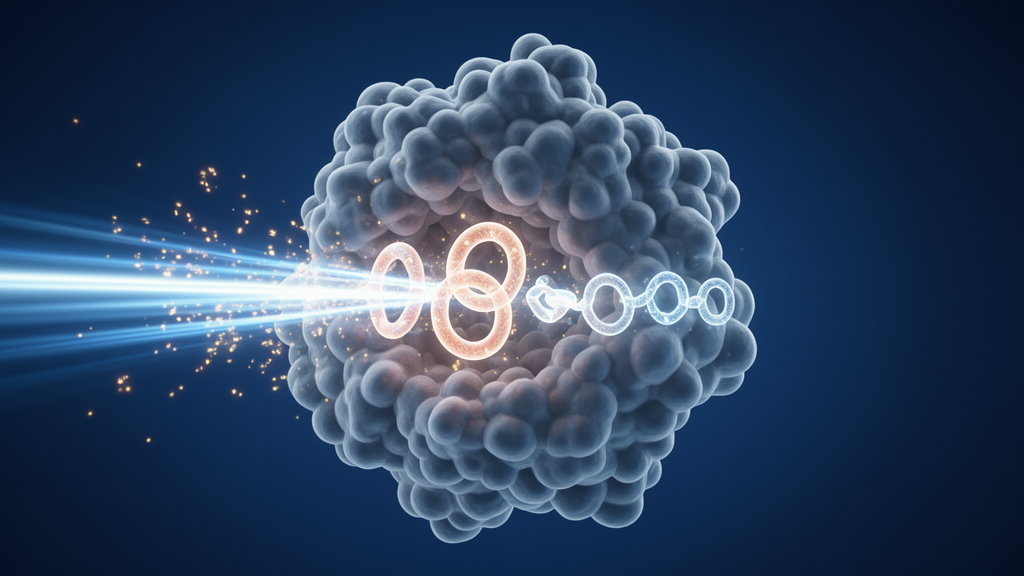
For more than half a century, scientists have known that the enzyme isopenicillin N synthase, or IPNS, performs one of the most remarkable feats of chemistry in biology: it converts a simple linear tripeptide into the strained, bicyclic ring system that gives penicillin its antibiotic power. Yet the fleeting molecular states through which the enzyme passes during this transformation have largely remained invisible. Now, using X-ray free-electron lasers, an international team of researchers has captured a sequence of transient intermediates in unprecedented detail, and in doing so has uncovered a step that no one anticipated: the sulfur atom of the substrate briefly lets go of the iron at the heart of the reaction.
IPNS belongs to the widespread family of Fe(II)-dependent oxygenases, enzymes that use iron and molecular oxygen to oxidize their substrates. Most members of this superfamily, which includes enzymes involved in collagen biosynthesis, lipid metabolism and DNA repair, carry out two-electron hydroxylation reactions coupled to the decarboxylation of a co-substrate. IPNS is unusual. It performs a four-electron oxidation, converting the tripeptide δ-(L-α-aminoadipoyl)-L-cysteinyl-D-valine, known as ACV, into isopenicillin N by forging two new rings: a four-membered β-lactam and a five-membered thiazolidine. Both ring-forming steps require the cleavage of strong carbon-hydrogen bonds, and both show large kinetic isotope effects when those hydrogens are replaced with deuterium.
Previous crystallographic work had established that ACV binds the catalytic iron through its cysteinyl sulfur atom, and spectroscopic and computational studies had proposed that oxygen binding generates a ferric superoxo species that abstracts a hydrogen from the cysteinyl β-carbon, producing a thioaldehyde intermediate and an iron(II)-hydroperoxide. But the actual structures of the intermediates involved in the ring-forming steps had never been observed directly. Two competing mechanisms had been proposed for how the O–O bond of the peroxide is cleaved and how the valine amide nitrogen is deprotonated, and the roles of water molecules in the active site were essentially unknown.
To resolve these questions, the team turned to time-resolved serial femtosecond crystallography, or tr-SFX, at X-ray free-electron laser facilities including the Linac Coherent Light Source in the United States, the Pohang Accelerator Laboratory XFEL in South Korea and SACLA in Japan. XFEL pulses are so brief and so intense that diffraction data can be collected from tiny microcrystals before radiation damage destroys the sample, making the method ideal for studying radiation-sensitive high-valent iron intermediates. The researchers grew anaerobic microcrystals of the IPNS:Fe(II):ACV complex, roughly three by three by eighty micrometres in size, and delivered them on a conveyor tape using acoustic droplet ejection. Nanolitre droplets were passed through a chamber filled with pure oxygen, and the reaction time was controlled simply by varying the speed of the tape between oxygen exposure and the X-ray pulse.
A clever isotope trick helped the team trap the most elusive intermediate. Because the second hydrogen abstraction step, at the valine carbon, exhibits a very large kinetic isotope effect of roughly thirty, the researchers used a deuterium-labelled version of the substrate, ACdV, in which the valine hydrogen is replaced with deuterium. This dramatically slows the second ring-forming step, allowing the monocyclic β-lactam intermediate to accumulate. The team collected structural data at one, two, three, four, six, eight and ten seconds of oxygen exposure, deliberately randomizing the order of data collection and interspersing water-only control runs to eliminate bias, and repeated all experiments at least twice.
The first two seconds revealed what was expected: an oxygen molecule binding end-on to the iron at the site trans to Asp216, with the occupancy of the bound oxygen rising from about fifty percent at one second to more than ninety percent at two seconds. Time-resolved X-ray emission spectroscopy, collected simultaneously from the same crystals and pulses, confirmed the formation of the Fe(III)-superoxo species. Then came the surprise. At three, four and six seconds, the electron density showed that the ACV sulfur atom had moved away from the iron, with the iron–sulfur distance stretching from about 2.3 angstroms to roughly 4.2 angstroms. The density was consistent with a thioaldehyde whose cysteinyl α-carbon retained its tetrahedral geometry, meaning the thioaldehyde did not coordinate the iron at all, contrary to what many mechanistic models had assumed.
The X-ray emission spectra at these timepoints showed the iron had been reduced back to Fe(II), consistent with the proposed iron(II)-hydroperoxide state. By eight and ten seconds, the sulfur had returned to the iron, restoring the Fe–S bond at about 2.4 angstroms, and the structures revealed the monocyclic β-lactam intermediate at seventy-five percent occupancy. This intermediate closely matched a previously determined structure of IPNS bound to a substrate analogue that stalls at the same stage, providing strong independent support for the assignment. Notably, at the monocyclic intermediate stage the valine hydrogen now points toward the iron, positioned productively for the second abstraction, whereas in the anaerobic starting structure it points away. The valine isopropyl group, which had been disordered in earlier timepoints, became well ordered, reflecting the conformational rigidity conferred by the restored iron–sulfur linkage.
The refined iron–oxygen bond at these late timepoints came out at about 1.96 angstroms, somewhat longer than the roughly 1.64 angstroms expected for a classic Fe(IV)=O species, though the interpretation is complicated by residual peroxide from the thioaldehyde population. To rule out artefacts, the team also determined a structure of IPNS complexed with a catalytically inactive vanadyl ion, V(IV)=O, which mimics the geometry of a high-valent metal-oxo species. The vanadium–oxygen bond refined to 1.64 angstroms, matching literature values and indicating that the elongated iron–oxygen distance was not a radiation-damage artefact. The spectroscopic data at eight and ten seconds showed the iron in a state more oxidized than Fe(III), supporting at least partial presence of the Fe(IV) intermediate critical for the second hydrogen abstraction.
Beyond the iron coordination sphere itself, the study revealed that water molecules and distant parts of the protein play decisive roles. B-factor analysis, a measure of atomic mobility, showed that the α3 and α10 helices become progressively more mobile during catalysis, with α3 returning to its starting conformation in the product complex while α10 remains dynamic, consistent with the movement needed to release the product. A chain of three water molecules extending from the iron-bound water W1 through W2 and W3 proved essential. Mutating the second-shell residue Asn252, which hydrogen-bonds to W2, produced a striking result: the conservative N252D substitution, which preserved the water chain structurally, abolished penicillin production in solution and instead yielded a hydrated aldehyde shunt product, while the bulkier N252Q and N252E variants disrupted W2 binding and impaired catalysis. A separate variant, H270E, in which the iron-coordinating His270 was replaced with glutamate, displaced the iron-bound water W1 and stalled the reaction at the thioaldehyde stage, with the released intermediate tautomerizing to enethiol products identified by comparison with synthetic standards.
Quantum mechanics/molecular mechanics calculations tied the observations together, supporting a pathway in which the iron-bound water W1 mediates peroxide cleavage to generate the Fe(IV)=O species, followed by water-assisted proton transfer to form the monocyclic intermediate. The computations reproduced the transient elongation of the iron–sulfur bond at the thioaldehyde stage and the low barrier for the final radical recombination that delivers isopenicillin N in a strongly exothermic step. Together, the structural, spectroscopic, mutagenic and computational evidence paints a revised picture of penicillin biosynthesis in which protein dynamics, a delicate water network and an unexpected release of sulfur from the iron choreograph the formation of one of medicine’s most important molecular scaffolds. The findings not only illuminate IPNS itself but offer a framework for understanding the broader Fe(II) oxygenase superfamily, and they demonstrate the power of X-ray free-electron laser methods to resolve transient enzymatic intermediates that no other experimental or computational approach can currently capture at such resolution.
Subject of Research: Time-resolved structural studies of intermediates in isopenicillin N synthase catalysis during β-lactam antibiotic biosynthesis
Article Title: Unanticipated intermediates during isopenicillin N synthase catalysis identified by time-resolved X-ray free-electron laser studies
Article References: Rabe, P., Makita, H., Southwart, R., Thomas, M. G., Kazaks, M., Basak, S., Zhou, T., Myers, W. K., Doyle, M. D., Simon, P. S., Hayama, S., Mosselmans, J. F. W., Stead, A. T., Oluwole, A. O., Sami, M., Clifton, I., Aller, P., Bhowmick, A., Tumber, A., … Schofield, C. J. (2026). Unanticipated intermediates during isopenicillin N synthase catalysis identified by time-resolved X-ray free-electron laser studies. Nature Catalysis. https://doi.org/10.1038/s41929-026-01618-4
Image Credits: AI Generated
DOI: 10.1038/s41929-026-01618-4
Keywords: isopenicillin N synthase, penicillin biosynthesis, X-ray free-electron laser, serial femtosecond crystallography, enzyme mechanism, iron oxygenase, β-lactam antibiotics, thioaldehyde intermediate, X-ray emission spectroscopy, protein dynamics, active-site water, structural biology
News Source: Bethany Barker. (October 9, 2026). X-ray Lasers Catch Penicillin-Making Enzyme in the Act, Revealing Surprise Intermediates. Scienmag.