
FRANKFURT — An international collaboration has shed light on the rapid transformations experienced by biologically significant molecules under the influence of ultraviolet (UV) radiation. This groundbreaking research, involving teams from Goethe University Frankfurt, the European XFEL in Schenefeld, and the Deutschen Elektronen-Synchrotron DESY in Hamburg, has unveiled the nuances of these ultra-fast processes, making them perceptible through innovative X-ray imaging techniques. The implications of this study extend far beyond understanding molecular behavior; they promise to revolutionize how scientists analyze various complex biomolecules.
At the center of this study is the molecule 2-thiouracil, belonging to a class of pharmaceutical compounds that utilize specific DNA building blocks known as nucleobases. Markus Gühr, head of the free-electron laser FLASH at DESY and a professor of chemistry at the University of Hamburg, indicates that 2-thiouracil contains a unique sulfur atom. This atom grants the molecule medically significant properties; however, it also increases its reactivity when exposed to UV radiation—a concern that ties this chemical behavior to potential skin cancer risks.
The research team employed a novel method, taking advantage of Coulomb explosion imaging. This technique uses intense X-ray pulses to knock electrons out of molecules. The resulting positive charge destabilizes the molecular structure, causing it to fragment almost instantaneously. Understanding the directions in which these fragments disperse allows researchers to infer the original configuration of the molecule. This scientific endeavor transcends previous limitations that restricted Coulomb explosion imaging to only the simplest molecular structures.
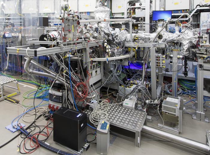
The research team masterfully merged this imaging technique with the extraordinary capabilities of the European XFEL’s SQS (“Small Quantum Systems”) instrument, which is recognized as the world’s most powerful X-ray laser. Michael Meyer, head of the SQS instrument, describes the experiment as a technical leap forward. For the first time, imaging techniques can be applied to biologically significant molecules like 2-thiouracil, marking a pivotal point in the intersection of fundamental physics and medical research.
To conduct the experiment, the researchers passed individual molecules through a fine gas nozzle into the X-ray beam, ensuring that only isolated molecules were targeted. This precision allowed them to focus the experimental setup and heightened the clarity of their observations. An additional UV pulse was delivered just prior to the X-ray pulse, effectively priming the molecules and enabling the observation of their structural changes as they responded to stimulation.
In exploring the time dynamics of these molecular transformations, the research team discovered that by altering the interval between the UV and X-ray pulses, they could effectively create a “slow-motion” video of the molecular events unfolding at an astonishing speed of 100 to 1000 femtoseconds, a time scale that challenges contemporary observational methodologies. Utilizing a sophisticated detector system, the researchers tracked the impact points of various atoms, rendering their findings palpable.
The analysis pointed to two pivotal discoveries about the 2-thiouracil molecule itself. The first significant revelation is related to the structural changes prompted by UV radiation. The normally flat 2-thiouracil molecule undergoes bending due to UV exposure, resulting in the sulfur atom protruding from its structure—an unstable configuration that results in heightened chemical reactivity. This reactivity not only distinguishes 2-thiouracil from other similar nucleobases but also raises substantial concerns regarding its potential health impacts.
In contrast, standard nucleobases are equipped with mechanisms to dissipate UV radiation harmlessly as heat, protecting them from potential damage. The unique configuration of 2-thiouracil, characterized by its sulfur atom, highlights a critical pathway towards understanding the associated risks of such molecules under UV light exposure. This fundamental knowledge opens avenues for developing drugs with greater efficacy and safety profiles.
The second major finding revolves around the capabilities of Coulomb explosion imaging itself. The researchers determined that they did not need to meticulously track every atom within the molecule to reconstruct its structure and analyze the changes it underwent. By concentrating on the sulfur and oxygen atoms and the four hydrogen nuclei, they successfully bypassed the cumbersome task of accounting for the six carbon atoms, simplifying future measurements of more complex molecular landscapes.
As this research progresses, it becomes increasingly evident that the scientific community stands on the brink of a transformative era in molecular analysis. The innovative techniques developed in this study extend far beyond the current horizon of molecular imaging, offering researchers new tools to dissect complex biological phenomena. The implications are vast, ranging from fundamental chemistry to applied drug design and therapeutic interventions.
By showcasing the merits of merging avant-garde techniques with traditional biological inquiry, this research not only clarifies the actions occurring at the molecular level but inevitably impacts how researchers and clinicians should view the relationship between UV exposure and molecular health outcomes. The ongoing dialogue surrounding drug safety, cancer risk, and molecular behavior will surely be enriched by these insights.
In summary, the study of 2-thiouracil as illuminated by this research not only enhances our understanding of molecular changes triggered by UV light but paves the way for future breakthroughs in both pharmacology and cancer research. The unique intersection of advanced X-ray techniques and biological inquiry may indeed revolutionize how we understand molecular interactions, providing novel insights that can lead to innovative approaches to drug development and cancer prevention.
Subject of Research: Chemical reactivity and structure of 2-thiouracil under UV radiation.
Article Title: Direct observation of ultrafast symmetry reduction during internal conversion of 2-thiouracil using Coulomb explosion imaging.
News Publication Date: 28-Feb-2025.
Web References: DOI: 10.1038/s41467-025-57083-3.
References: Not applicable.
Image Credits: Credit: European XFEL.
Keywords: 2-thiouracil, Coulomb explosion imaging, molecular imaging, UV radiation, skin cancer, X-ray laser, molecular structure, pharmacology, drug development, chemical reactivity, biochemistry, molecular transformations.
Tags: 2-thiouracil analysisActive substances in chemistryBiologically significant moleculesComplex biomolecules analysisCoulomb explosion imagingInnovative research collaborationsMolecular transformations studyNucleobases in DNAPharmaceutical compounds researchSkin cancer risk factorsUltraviolet radiation effectsX-Ray imaging techniques

