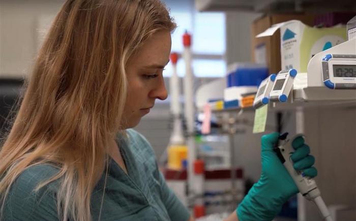
West Virginia University researchers are now able to view synthetic DNA at the atomic level, giving them the ability to understand how to change its structure in hopes of enhancing its scissor-like function. Learning more about these synthetic DNA reactions could be the key to unlocking new technology for medical diagnoses and treatments.

Credit: WVU Photo/Hannah Maxwell
West Virginia University researchers are now able to view synthetic DNA at the atomic level, giving them the ability to understand how to change its structure in hopes of enhancing its scissor-like function. Learning more about these synthetic DNA reactions could be the key to unlocking new technology for medical diagnoses and treatments.
In the chemistry world, the findings help answer a 30-year-old question about this specific DNA structure and how scientists can get it to produce a reaction without changing the DNA itself, a process called catalysis.
“This is only, maybe, the third example lending insights, at the very detailed atomic level, into how chemically active DNA promote their unique functions that give all these applications their power,” said Aaron Robart, associate professor in the WVU School of Medicine Department of Biochemistry and Molecular Medicine, and principal investigator of the project, funded by a competitive Ralph E. Powe Junior Faculty Enhancement Award. “Atomic detail gives us a long-sought road map to start building and improving a technology that can be broadly applicable to health and diagnostics.”
Robart said once scientists understand how to make the technology function more efficiently, it could theoretically be applied as treatment for diseases such as retinal degeneration or cancer.
The researchers’ findings are published in Communications Chemistry, a Nature family journal.
Robart points out synthetic DNA used in the study, known as DNAzymes, is different from human DNA. Created in a lab, DNAzymes are inexpensive to produce and capable of catalyzing chemical reactions. They’ve been artificially evolved to perform such functions as monitor air quality and measure heavy metals that have leached into soil.
“Typically, we think of DNA as inert, serving as a storage unit for our genetic information,” Robart said. “However, there are certain types of DNA evolved in the laboratory that defy the conventional rules. These DNAs can fold into complex shapes, enabling them to perform a remarkable range of reactions.
“The only problem is, after 30 years of research, we really didn’t have a clue on how any of the chemistry was happening. One of the big things we were missing is what our lab does with crystals, resulting in high resolution structures of what nucleic acids look like down to the atom detail and how they can do all this chemistry.”
To be able to see DNA at the atomic level, Robart and his lab students, Evan Cramer, of Lake Ann, Michigan, Sarah Starcovic, of Cameron, and Beka Avey, of Martinsburg, collaborated with Advanced Photon Source at the U.S. Department of Energy’s Argonne National Laboratory in Chicago. The process — X-ray crystallography — involves crystallizing synthetic DNA and then zapping it with super powered X-rays to reveal its structure. Working with APS, the team was able to control the X-ray and collect data via the internet.
“Using this information, we can better understand how other DNAzymes may behave in their cleavage reactions,” said Starcovic who is pursuing a doctoral degree in biochemistry and molecular medicine.
Robart said what they saw was a structure with little arms that can reach out to find another section of a complementary sequence and clamp themselves together, similar to the way Velcro attaches.
“These DNAs can act as molecular scissors with precise specificity to cut RNA or DNA, or they can function as glue,” Robart explained. “Say you have a mutated gene that’s causing disease, we could get this DNA into the cells and it would be able to get rid of all that kind of message that’s causing the proteins that lead to the disease.”
Cramer, lead author of the published paper and a biochemistry and molecular medicine doctoral student, said he hopes future studies fill knowledge gaps for clinical implementation.
“It is difficult to improve something when how it works is not entirely known,” he said.
A recent Ruby Scholars Graduate Fellow, he’ll continue on the research team with a BridgesDH NSF-NRT Fellowship.
Robart said the next step is to focus on alternative techniques for capturing DNAzymes at different points along their function.
“It will be like we’re making an old school animation molecular flipbook,” Robart said. “This level of detail is used to understand how to improve, target and regulate their activity. This is only one of hundreds of different varieties of DNAzymes, all with their own unique properties begging to be applied to topics in human health.”
He said he also hopes to gain insight from School of Medicine colleagues on how the model systems could be used for therapeutics.
“We’re in a unique spot,” Robart said. “We have a potential cure in search of a disease. I feel fortunate to be in an environment surrounded by so many talented collaborators in the School of Medicine to help this exciting technology reach its full potential.”
Journal
Communications Chemistry
DOI
10.1038/s42004-023-00924-3
Article Title
Structure of a 10-23 deoxyribozyme exhibiting a homodimer conformation
Article Publication Date
10-Jun-2023


