Researchers have mapped in detail how human urine-derived cells can be reprogrammed into induced presomitic mesoderm (UiPSM), a laboratory-made counterpart of the embryonic tissue that gives rise to vertebrae, skeletal muscle, and cartilage. In a study published in Cell Regeneration, a team led by Kaipeng Wang and Yue Qin combined transcriptomic, chromatin accessibility, and functional analyses to dissect the roles of two key components of the induction system: the Wnt activator CHIR99021 and the Dot1L inhibitor EPZ5676. Their central finding is that these two compounds play strikingly different jobs. CHIR-driven Wnt/β-catenin signaling is indispensable for triggering the fate conversion, while EPZ5676 controls when reprogramming happens and how efficiently it proceeds, without dictating the final cell identity.
The UiPSM system itself was established in earlier work by the same group. Urine-derived cells are plentiful, easy to obtain, and non-invasive, which makes them an attractive starting material for generating patient-specific models of human somitogenesis. The presomitic mesoderm (PSM) is the cellular basis of somite formation, and its dysregulation in human development is linked to congenital scoliosis and vertebral malformations. Because ethical constraints limit direct study of human embryos, and rodent models differ in important ways from human development, an in vitro system built from human somatic cells offers a valuable alternative window into mesoderm lineage specification.
To probe the chemistry of the reprogramming cocktail, the team systematically removed individual components from the induction medium, which contained CHIR99021, EPZ5676, basic fibroblast growth factor (bFGF), and epidermal growth factor (EGF). Removing CHIR completely abolished UiPSM colony formation. PSM marker genes such as T, TBX6, MIXL1, and CDX2 failed to be upregulated, while urinary cell markers PAX8 and CD13 stayed highly expressed. In contrast, withdrawing either bFGF or EGF had no discernible effect, and did not redirect cells toward lateral or intermediate mesoderm fates. Flow cytometry of T-positive cells quantified the difference: CHIR withdrawal nearly eliminated induction efficiency, dropping T-positive cells to about 4.5 percent, while EPZ removal cut efficiency roughly in half, from 52.5 percent to 27.5 percent.
RNA sequencing across days 0, 3, 6, and 9 of induction sharpened the picture. In the complete induction condition, biological processes tied to somite development, somitogenesis, and anterior–posterior patterning were enriched on days 6 and 9. In the EPZ-withdrawal group, these processes appeared only on day 9, indicating a pronounced delay rather than a failure. Genes normally activated by day 6 were not switched on until day 9 without EPZ. In the CHIR-withdrawal group, by contrast, these developmental programs were never enriched at any time point, and representative genes including WNT3A, TBX6, FGF8, CDX2, DLL1, NOTCH1, and HES7 were almost completely abolished. The authors conclude that CHIR-mediated Wnt signaling is indispensable for UiPSM reprogramming, whereas EPZ5676 facilitates the timely induction of PSM-associated genes.
Chromatin-level analysis using ATAC-seq revealed how the two compounds differ mechanistically. Comparing accessibility profiles between human urine-derived cells and PSM cells, the researchers classified loci into three categories: closed in urine cells and open in PSM cells (CO), open in urine cells and closed in PSM cells (OC), and permanently open in both (PO). CHIR withdrawal disrupted the reorganization of both CO and OC loci, preventing proper chromatin remodeling. Roughly 60 percent of loci failed to open properly and about 90 percent failed to close correctly without CHIR, compared with approximately 20 percent and 30 percent, respectively, without EPZ. EPZ withdrawal largely preserved the overall remodeling pattern but reduced the intensity of accessibility signals and delayed the timing of chromatin-opening events, arresting chromatin maturation at an intermediate stage.
Motif analysis of the CHIR-dependent open regions pointed to a specific set of transcription factors. The CO loci were enriched for binding motifs of the paraxial mesoderm factors T (Brachyury), CDX2, and TBX6, alongside T-box, HMG, Forkhead, and Homeobox motifs. The team then tested whether overexpressing nine candidate factors, collectively termed 9F, could substitute for CHIR. Overexpression of the nine-factor panel in CHIR-free medium generated a limited number of colonies, designated 9F-UiPSM, which partially restored endogenous T, TBX6, and CDX2 expression and could be passaged at least five generations while maintaining SALL4 and SOX2, markers of neuromesodermal progenitor stemness. Notably, overexpressing 9F in the absence of EPZ did not change colony numbers, indicating that these factors do not mediate EPZ’s effects.
A pairwise combinatorial screen narrowed the field further. Among the six factors that were actually expressed during reprogramming, the combinations T/CDX2 and CDX2/TBX6 showed the highest efficiency in promoting UiPSM induction without CHIR, producing colonies designated CT-UiPSM and C6-UiPSM that maintained stable morphology and SOX2/SALL4 expression through passage five. ATAC-seq correlation analysis showed that these factor-induced clones closely resembled standard UiPSM clones and were clearly distinct from the parental urine cells. When differentiated into self-organizing human somitoids, the C6 and CT groups reached formation efficiencies of 59 and 63 percent respectively, compared with roughly 85 percent for standard UiPSM-derived somitoids, while the 9F group managed only 11 percent. Elongation rates among the resulting structures did not differ significantly. The poor performance of the full nine-factor cocktail appears to stem from inhibitory effects of FOXA1 and FOXA2, since deleting both factors improved colony formation and endogenous CDX2 and FGF8 expression.
The study also uncovered a positive feedback loop that sustains UiPSM identity. CDX2 emerged as a key regulator because its expression is induced early during reprogramming and increases with CHIR concentration. Generating a CDX2-knockout UiPSM line using CRISPR-Cas9, the researchers found that loss of CDX2 disrupted PSM identity and impaired anterior–posterior axis elongation in somitoid differentiation, with similar results from knockdown experiments. CUT&Tag and ATAC-seq analyses showed that CDX2 directly binds regulatory elements of WNT3A, T, and TBX6. Knocking out CDX2 markedly reduced chromatin accessibility and H3K27ac enhancer marks at these loci, almost completely abolished WNT3A transcription, substantially downregulated T, and reduced TBX6 expression. WNT3A loss was accompanied by reduced total and active β-catenin levels and increased AXIN2, a negative regulator of Wnt signaling. Overexpressing WNT3A or WNT5A partially compensated for CHIR absence, with WNT3A exerting the stronger effect, though neither fully recapitulated CHIR’s reprogramming effects.
Knockdown experiments among the three core factors revealed a mutually interdependent network. CDX2 knockdown affected both T and TBX6, TBX6 knockdown altered CDX2 and T, and T knockdown primarily affected CDX2, with all three perturbations influencing MSGN1, a PSM marker. Together with inputs from Wnt/β-catenin signaling, these interactions form a dynamically balanced regulatory network in which CDX2 drives WNT3A expression, WNT3A reinforces β-catenin activity, and β-catenin sustains the core transcription factors. Separately, the team found that exogenous FGF agonists were largely redundant, but inhibiting endogenous FGF signaling with the receptor inhibitor PD173074 reduced UiPSM marker gene expression and impaired axial elongation, suggesting that endogenous FGF production suffices to support UiPSM development.
The authors are careful to note the limits of their conclusions. They state that the molecular regulatory cascades are supported mainly by correlative multi-omic profiling and small-molecule interventions, and that definitive causal relationships require further validation through gene knockout and overexpression functional assays. They also flag unresolved questions, including the downstream epigenetic effects of EPZ5676, the individual roles of each factor in the nine-factor panel, and the possible involvement of non-canonical Wnt pathways such as planar cell polarity signaling. Even so, the work offers a simplified two-factor route to UiPSM induction through CDX2/T and CDX2/TBX6, and strengthens UiPSM as a human cellular platform for studying congenital scoliosis, vertebral malformations, and mesoderm-derived tissue regeneration.
Subject of Research: Mechanisms of Wnt signaling and EPZ5676 in reprogramming human urine-derived cells into induced presomitic mesoderm
Article Title: Distinct regulation of Wnt signaling and EPZ5676 governs human UiPSM reprogramming via the CDX2/T/TBX6 core axis
Article References: Wang, K., Huang, X., Gao, H., Li, J., Huang, T., Ke, M., Fu, Y., Lin, Z., Pei, D., & Qin, Y. (2026). Distinct regulation of Wnt signaling and EPZ5676 governs human UiPSM reprogramming via the CDX2/T/TBX6 core axis. Cell Regeneration, 15(1), Article 28. https://doi.org/10.1186/s13619-026-00302-z
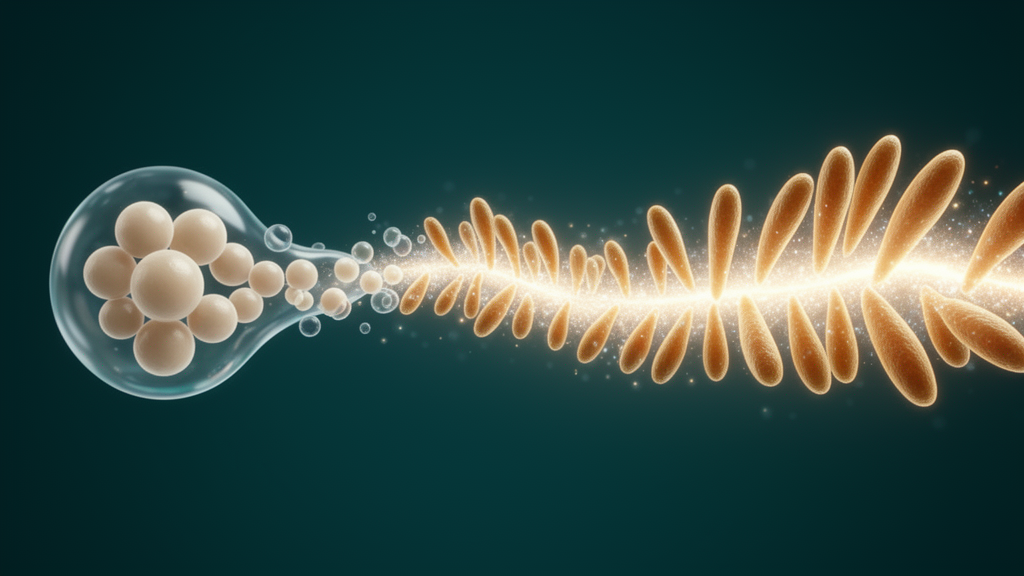
Image Credits: AI Generated
DOI: 10.1186/s13619-026-00302-z
Keywords: UiPSM, presomitic mesoderm, Wnt signaling, CHIR99021, EPZ5676, CDX2, Brachyury, TBX6, chromatin remodeling, somitogenesis, cell reprogramming, WNT3A
News Source: Gregory Coleman. (October 28, 2026). Wnt Signaling and CDX2/T/TBX6 Axis Drive Human Urine Cell Reprogramming into Presomitic Mesoderm. Scienmag.