


Wilson’s disease is famous in neurology textbooks for a fairly predictable signature: liver trouble paired with symmetric abnormalities deep in the basal ganglia on brain MRI. A new case report from Children’s Hospital of Nanjing Medical University, published in Frontiers in Pediatrics, describes a 13-year-old boy whose illness broke that pattern almost completely. His scans showed extensive, frontally dominant white matter disease that initially led clinicians to suspect Alexander disease, a rare inherited leukodystrophy. Only after a broad diagnostic work-up, including whole-exome sequencing, did the true diagnosis emerge: a homozygous pathogenic mutation in the ATP7B gene, the genetic cause of Wilson’s disease.
The boy’s story began subtly. Over the course of a year, he experienced insidious speech deterioration, drooling, declining academic performance, and social withdrawal. His symptoms were initially attributed to behavioral issues, and it was only when he developed intermittent twitching around the mouth that his family sought medical attention. By the time he was examined at age 13 years and 6 months, neurological testing revealed dysarthria, bradykinesia, mild lower limb spasticity, hyperreflexia, and a short-stepped gait. Notably, his liver transaminases and other routine laboratory tests were within normal limits, which made an underlying copper-metabolism disorder far less obvious.
There was, however, one retrospective clue. His parents later recalled an episode of acute hepatitis at age 5 to 6 years, with alanine aminotransferase levels exceeding 1,000 U/L, which had resolved completely without an identified cause. The report’s authors suggest that this early liver injury may have represented the initial hepatic phase of Wilson’s disease, and that subsequent normalization of transaminases could reflect mild early liver injury, the self-repair capacity of young hepatocytes, or even the resolution of a concurrent viral infection masking the persistent underlying pathology.
Additional work-up at presentation hinted at multiorgan involvement. Urinalysis showed occult blood, proteinuria, and a red blood cell count of 111.7 per microliter with mixed morphology, and urinary tract ultrasound indicated bilateral renal parenchymal enhancement. Video electroencephalography recorded bilateral frontal pole, frontal, and midline spike waves, spike-and-wave complexes, and rhythmic discharges, predominantly on the left side, along with two focal electrical seizures arising from the left frontal region. Combined with the intermittent perioral twitching, these findings raised suspicion of focal epileptic seizures. The care team recommended oral antiepileptic drugs, but the parents declined.
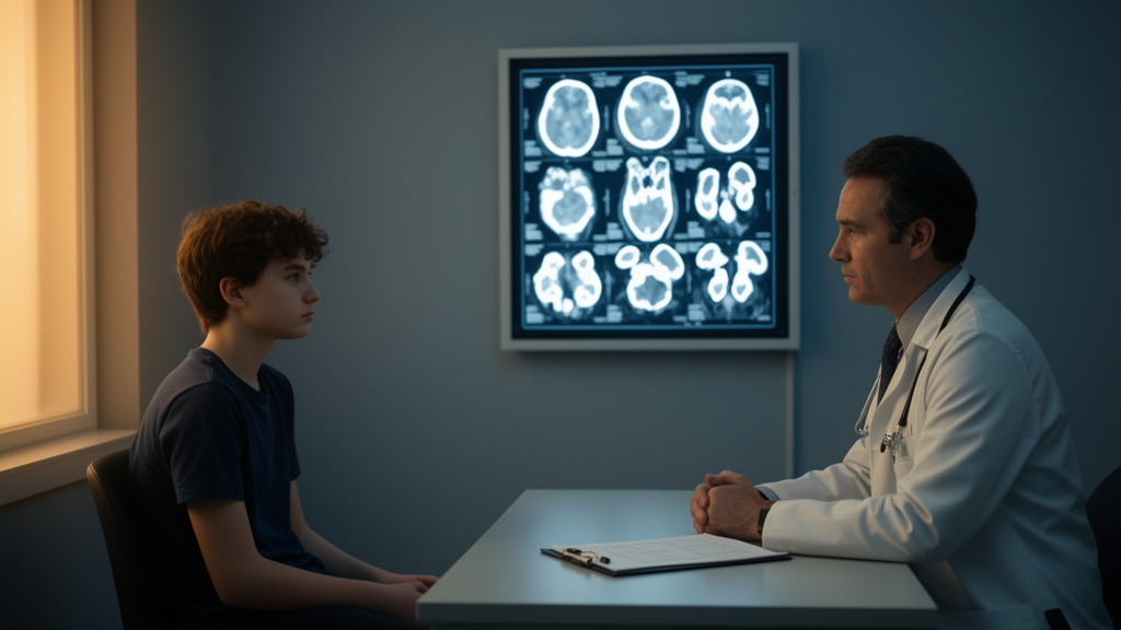
The neuroimaging was the most striking element of the case. Non-contrast computed tomography revealed bilateral hypodense areas in the frontal lobes. Brain MRI performed on a 3.0 Tesla scanner then demonstrated multifocal, bilateral, and symmetric abnormalities: extensive T2-weighted and FLAIR hyperintense signal in the periventricular and deep frontal white matter, extending into the corpus callosum, basal ganglia, thalamus, midbrain, and pons. Within the frontal white matter, several foci showed central signal loss on FLAIR, indicating cystic encephalomalacia—softened, fluid-filled tissue where brain substance had been lost. Diffusion-weighted imaging additionally showed patchy restricted diffusion in the frontal cortical and subcortical areas and in the basal ganglia, and volumetric analysis disclosed mild atrophy of the basal ganglia.
Given the frontal-predominant leukoencephalopathy with brainstem involvement, Alexander disease was considered the primary radiological differential diagnosis, with metachromatic leukodystrophy and adrenoleukodystrophy also in the frame. This pattern matters because classic neurological Wilson’s disease typically produces symmetric T2 hyperintensity in the basal ganglia, thalamus, and brainstem, not diffuse frontal white matter disease. Frontal-dominant leukoencephalopathy in Wilson’s disease has been reported in the literature but is rare in clinical practice, and white matter disease accompanied by cystic change is rarer still. The authors argue that the coexistence of basal ganglia atrophy with white matter lesions, and cystic change suggesting chronic progression or severity, may serve as critical diagnostic clues pointing back toward Wilson’s disease.
The restricted diffusion on DWI adds another layer of interest. The report notes that cortical DWI hyperintensity is relatively uncommon in Wilson’s disease, citing a prior hypothesis that DWI hyperintensity in the basal ganglia of affected patients results from copper overload-induced cellular damage, driving inflammation, cellular swelling, and ultimately cell death. The authors suggest the multiple cortical hyperintensities in this case may reflect cytotoxic edema or copper-associated inflammation, though this remains an interpretation of the imaging rather than a directly verified mechanism in this patient.
Diagnostic confirmation came from a combination of genetic and biochemical evidence. Serum ceruloplasmin was severely low at 0.0254 g/L against a reference range of 0.2 to 0.6 g/L, 24-hour urinary copper excretion was elevated at 122.0 micrograms against a reference range of 15 to 60 micrograms, and slit-lamp examination confirmed the presence of Kayser-Fleischer rings, the copper deposits in the corneal rim that are a hallmark of the disease. Using the Leipzig scoring system, the patient scored 10 points, confirming the diagnosis. Treatment with a low-copper diet, penicillamine, and zinc sulfate was initiated.
The follow-up story is a cautionary one. The patient was transferred to outpatient care largely because the family lived far from the treatment center, and the parents, having initially declined antiepileptic medication, later reported by telephone that the boy’s clinical symptoms had improved without returning for further hospital evaluation. The report therefore documents the diagnosis and the start of chelation therapy, but not verified long-term neurological or hepatic outcomes—a limitation readers should keep in mind when interpreting the case.
Why does this single patient matter beyond his own chart? Wilson’s disease is an autosomal recessive disorder of copper metabolism caused by biallelic pathogenic variants in ATP7B, whose protein product mediates copper incorporation into ceruloplasmin and biliary copper excretion; when that function is lost, copper accumulates toxically in the liver and then elsewhere, producing multiorgan injury. It is, as the authors emphasize, a treatable neurogenetic disorder: with timely diagnosis, lifelong individualized therapy aimed at a negative copper balance, symptomatic medications for tremor and dystonia, and rehabilitation, long-term survival in fully treated and compliant patients appears similar to the general population. The corollary is that diagnostic delay carries real cost. The authors argue that etiological evaluation of unexplained hepatitis in preschool-aged children, including early measurement of serum ceruloplasmin and urinary copper, could have prevented the irreversible neurological dysfunction in this case, and they recommend that Wilson’s disease be included in the differential diagnosis of pediatric progressive bilateral frontal leukoencephalopathy—even when classic hepatic findings or pronounced extrapyramidal signs are absent, and even when the scans look for all the world like an inherited leukodystrophy.
Subject of Research: A pediatric case report of Wilson's disease presenting as frontal-dominant leukoencephalopathy mimicking Alexander disease.
Article Title: A case report of Wilson's disease with rare frontal lobe white matter lesions
Article References: Du, X., Guo, H., Ding, L., Liang, C., Zhang, G., & Ginowree, N. B. N. (2026). A case report of Wilson's disease with rare frontal lobe white matter lesions. Frontiers in Pediatrics, 14, Article 1874872. https://doi.org/10.3389/fped.2026.1874872
Image Credits: AI Generated
DOI: 10.3389/fped.2026.1874872
Keywords: Wilson's disease, ATP7B, leukoencephalopathy, Alexander disease, pediatric neurology, MRI, ceruloplasmin, Kayser-Fleischer rings, case report, copper metabolism, white matter, seizures
News Source: Ophelia Keating. (October 29, 2026). Wilson’s Disease Masqueraded as a Leukodystrophy in a Teenager. Scienmag.



