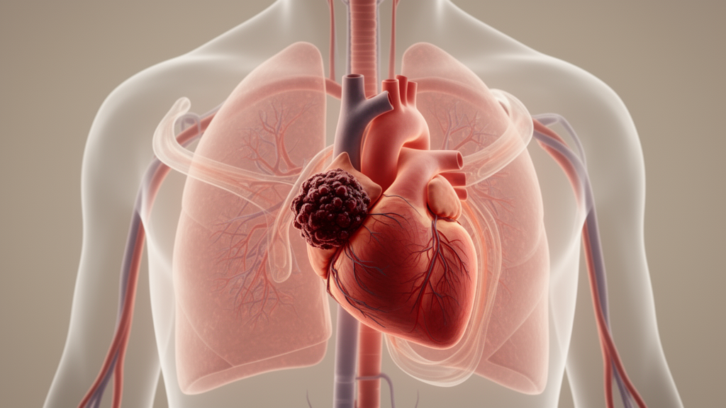
Patients with acute pulmonary thromboembolism (APTE) show different degrees of right ventricular strain depending on where in the leg their deep vein thrombosis (DVT) is located, according to a study published in BMC Cardiovascular Disorders. The finding, based on 231 first-time APTE patients, links more distal clot locations—those in the smaller leg veins—to higher levels of brain natriuretic peptide (BNP) and more pronounced echocardiographic markers of right ventricular load.
The research team, led by Feng Jiang and colleagues, categorized patients into four groups using lower limb vascular ultrasound: leg intermuscular vein thrombosis (94 patients), tibiofibular vein thrombosis (31), femoral popliteal vein thrombosis (90), and inferior iliac vein thrombosis (16). Patients were admitted between October 2013 and August 2020, and the study focused on first episodes of APTE.
The researchers compared clinical data, BNP levels, and several echocardiographic parameters across the four groups. These parameters included pulmonary artery systolic pressure (PASP), pulmonary artery trunk width, right ventricular outflow tract width, and right ventricular diameter. Baseline characteristics were reported as comparable across the groups, which strengthens the interpretation that differences in the measured outcomes relate to thrombosis location rather than underlying demographic differences.
The results showed that BNP levels, PASP, pulmonary artery trunk width, right ventricular outflow tract width, and right ventricular diameter were all significantly higher in the leg intermuscular vein thrombosis group and the tibiofibular vein thrombosis group than in the femoral popliteal vein thrombosis group and the inferior iliac vein thrombosis group, with statistical significance reported at P < 0.05.
Correlation analyses added a quantitative dimension to the comparison. The study found a significant positive correlation between DVT location and pulmonary artery trunk width (r = 0.654), right ventricular outflow tract width (r = 0.582), and right ventricular diameter (r = 0.741), all with P = 0.001. These correlation coefficients indicate moderately strong to strong associations between the anatomical position of the thrombus and the measured dimensions of the right-sided cardiac and pulmonary arterial structures.
Patients with distal DVT, meaning thrombosis in the leg veins, also exhibited a higher proportion of moderate to severe pulmonary hypertension. This observation suggests that the location of the clot within the venous system may carry information about the severity of the pulmonary vascular burden that follows an acute embolic event.
The authors conclude that the anatomical location of DVT, particularly leg vein thrombosis, is significantly associated with the degree of right ventricular strain after APTE. They further suggest that echocardiographic parameters and BNP levels may help stratify patients according to both DVT location and pulmonary hypertension severity.
The study’s design was observational and cross-sectional in its comparison of groups, and it included a relatively small number of patients in the inferior iliac vein thrombosis group (16 patients) and the tibiofibular vein thrombosis group (31 patients). The findings describe associations measured at the time of admission and do not establish causal mechanisms linking clot location to ventricular strain. The article was published as open access on 28 October 2026 and is shared as an accepted manuscript that remains subject to further editorial revision before the final version of record.
Subject of Research: The relationship between the anatomical location of lower extremity deep vein thrombosis and right ventricular strain after acute pulmonary thromboembolism.
Article Title: Association between anatomical location of lower extremity deep vein thrombosis and right ventricular strain parameters after acute pulmonary thromboembolism
Article References: Jiang, F., Sun, S., Pang, G., Zhao, Z., Mi, S., Zhang, Q., Liu, M., & Yu, G. (2026). Association between anatomical location of lower extremity deep vein thrombosis and right ventricular strain parameters after acute pulmonary thromboembolism. BMC Cardiovascular Disorders. https://doi.org/10.1186/s12872-026-06438-9
Image Credits: AI Generated
DOI: 10.1186/s12872-026-06438-9
Keywords: deep vein thrombosis, pulmonary thromboembolism, right ventricular strain, echocardiography, brain natriuretic peptide, pulmonary hypertension, pulmonary artery systolic pressure, vascular ultrasound, cardiovascular research, BMC Cardiovascular Disorders, Association, between
News Source: Ophelia Keating. (October 29, 2026). Where the Clot Sits May Shape Right Ventricular Strain After Pulmonary Embolism. Scienmag.