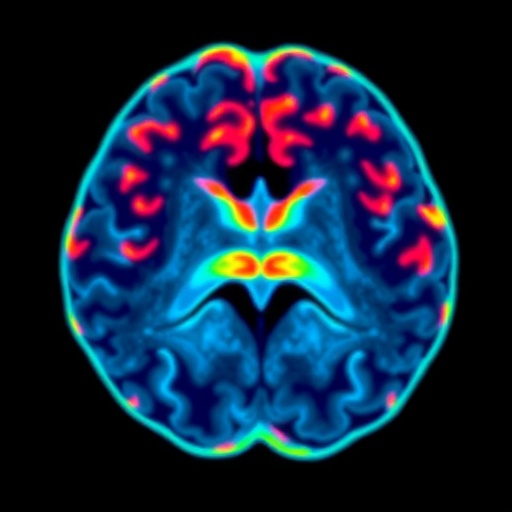
In a groundbreaking advancement in pediatric neuro-oncology imaging, researchers have unveiled a novel application of volumetric amide-proton-transfer weighted (APTw) magnetic resonance imaging (MRI) histogram analysis to distinguish between high-grade and low-grade gliomas in children. This innovative technique harnesses the subtle biochemical differences within tumor tissues, presenting a promising frontier for non-invasive diagnostic precision in pediatric brain tumors where differentiation is notoriously challenging.
Gliomas, accounting for the majority of pediatric brain tumors, present a wide spectrum spanning low-grade varieties that tend to grow slowly, to high-grade versions characterized by aggressive behavior and poor prognosis. Traditional imaging modalities often fall short in reliably distinguishing these grades due to overlapping morphological features, necessitating invasive biopsies that carry risks, especially in young patients. The study, spearheaded by Lin et al., innovatively capitalizes on the molecular sensitivity of APTw imaging, a technique that probes endogenous proteins and peptides via the exchange of amide protons, to offer a nuanced insight into tumor biology.
Conducted retrospectively on 69 pediatric patients suspected of possessing brain tumors, the study meticulously analyzed APTw imaging data accrued over a three-year period. From this cohort, 32 patients qualified for the final analysis after stringent inclusion criteria, encompassing a balanced distribution of males and females with a mean age of approximately 5.5 years. The researchers performed an extensive histogram evaluation focusing on two crucial tumor regions: the gross tumor core and the solid tumor components, extracting a diverse set of quantitative metrics that profile the heterogeneity and biochemical milieu within these neoplastic masses.
One of the most compelling aspects of the research lies in the statistical rigor applied. Recognizing the pitfalls of multiple comparisons, the team deployed Bonferroni correction to adjust significance thresholds, enhancing the robustness of their findings. Despite this conservative approach, certain histogram parameters demonstrated notable differences between pediatric low-grade gliomas (pLGG) and high-grade gliomas (pHGG). Specifically, within the gross tumor core, low-grade gliomas exhibited a higher minimum amide proton transfer value (APT_min), suggesting a distinct molecular signature compared to their high-grade counterparts.
Meanwhile, paradoxical patterns emerged in the solid tumor components where high-grade gliomas exhibited elevated maximum APT values (APT_max), higher variance, and greater entropy—statistical parameters indicating increased heterogeneity and complexity—compared to low-grade tumors. These findings underscore the biochemical diversity and chaotic microenvironment typical of aggressive tumor phenotypes. Interestingly, the minimum APT value in the solid components was lower for high-grade tumors, highlighting the complex and sometimes counterintuitive nature of tumor tissue characteristics captured by APTw imaging.
Further refinement of diagnostic accuracy was achieved through subgroup analysis comparing pilocytic astrocytoma—a common pediatric low-grade glioma variant—with high-grade gliomas. This comparison revealed that the 10th percentile APT value (APT 10th) from volumetric tumor data was significantly higher in pilocytic astrocytomas, providing a potent discriminatory marker. The diagnostic performance, quantified by the area under the receiver operating characteristic curve (AUC), reached an impressive 0.82, reflecting high sensitivity and specificity in distinguishing these tumor types.
The implications of this study are profound. Pediatric brain tumors require delicate therapeutic balancing acts, where accurate grading informs surgery extent, chemotherapy, and radiation planning. By introducing a non-invasive, quantitative imaging biomarker sensitive to tumor molecular composition, the use of APTw histogram analysis could minimize unnecessary surgical interventions and allow for more tailored therapies. This aligns with the broader precision medicine movement, which aims to customize medical care based on individual disease biology rather than purely anatomical criteria.
Moreover, the study highlights an intriguing divergence between pediatric and adult applications of APTw imaging. While the technique has shown promise in adult glioma differentiation, the unique pediatric tumor biology calls for specialized analytical frameworks, as evidenced by the distinct histogram metric patterns observed. Such nuances emphasize the necessity of age-appropriate imaging standards and validate the need for dedicated pediatric studies rather than extrapolating adult data.
Technically, the volumetric nature of APTw analysis underscores the advantage of three-dimensional tumor assessment over traditional two-dimensional slices. This holistic approach accounts for spatial heterogeneity and provides a comprehensive biochemical fingerprint of the tumor microenvironment, critical for understanding the complex biology of pediatric brain neoplasms. The study’s integration of rigorous imaging protocols with advanced statistical analysis sets a high methodological bar for future research in this domain.
The retrospective design warrants cautious interpretation, yet it establishes foundational knowledge and paves the way for prospective, multicenter trials with larger cohorts to verify and expand upon these findings. Additionally, integrating APTw imaging with other advanced modalities such as diffusion tensor imaging or perfusion MRI could further enhance diagnostic granularity, potentially unraveling tumor infiltration patterns and microvascular characteristics concomitantly.
From a clinical translation viewpoint, the widespread adoption of APTw histogram analysis hinges on the availability of compatible MRI protocols and post-processing tools, as well as training radiologists in interpreting these novel parameters. Collaborative efforts between imaging scientists, neurologists, and oncologists are imperative to standardize acquisition techniques, establish reference databases, and validate clinical utility through longitudinal studies assessing outcomes correlated with imaging biomarkers.
In conclusion, the study by Lin and colleagues represents a significant leap toward non-invasive characterization of pediatric gliomas, illuminating the path for molecular-level imaging biomarkers to guide diagnosis and management. By meticulously dissecting volumetric APTw histogram metrics, the researchers have unveiled a promising imaging signature that differentiates low-grade from high-grade pediatric gliomas with notable accuracy. This advancement not only enhances the neuroradiological toolkit but also resonates with the broader aspiration of personalized pediatric neuro-oncology care.
As technology progresses and molecular imaging becomes increasingly integral to clinical workflows, the impact of such innovations will reverberate throughout the landscape of pediatric brain tumor diagnosis and treatment. Future research inspired by these findings will likely focus on integrating metabolic, genetic, and immunological data with advanced imaging metrics, creating a multi-dimensional tumor profile that could revolutionize prognostication and therapeutic decision-making.
In essence, volumetric amide-proton-transfer weighted imaging histogram analysis stands poised to redefine pediatric glioma imaging. By moving beyond mere morphology toward intricate biochemical characterization, it offers hope for earlier, more precise, and less invasive diagnosis—a crucial advancement for improving outcomes in the vulnerable pediatric population struggling with brain tumors.
Subject of Research: Differentiation of pediatric high-grade and low-grade gliomas using volumetric amide-proton-transfer weighted (APTw) imaging histogram analysis.
Article Title: Volumetric amide-proton-transfer weighted imaging histogram analysis to differentiate pediatric high-grade and low-grade gliomas.
Article References:
Lin, G., Zhuang, Y., Lin, F. et al. Volumetric amide-proton-transfer weighted imaging histogram analysis to differentiate pediatric high-grade and low-grade gliomas. BMC Cancer 25, 1392 (2025). https://doi.org/10.1186/s12885-025-14822-5
Image Credits: Scienmag.com
DOI: https://doi.org/10.1186/s12885-025-14822-5
Tags: APTw imaging techniquebiochemical differences in tumorsendogenous proteins and peptides in MRIglioma differentiation in childrenhigh-grade vs low-grade gliomasmagnetic resonance imaging histogram analysisnon-invasive brain tumor diagnosispediatric brain tumor classificationpediatric neuro-oncology imagingretrospective study on pediatric patientstraditional imaging limitationsvolumetric amide-proton transfer imaging


