In a pioneering advancement that could reshape the landscape of molecular biology and biomedical imaging, researchers from Albert Einstein College of Medicine and the Salk Institute for Biological Studies have unveiled a transformative fluorescent probe technology, poised to significantly enhance the precision with which scientists visualize proteins inside living cells and tissues. Published in the latest issue of Nature Methods, this breakthrough employs engineered fluorescent nanobodies—compact, antibody-derived protein fragments—that fluoresce exclusively upon binding their designated targets. This on-demand activation system presents a quantum leap in imaging fidelity, overcoming long-standing challenges posed by background noise inherent in conventional fluorescent probes.
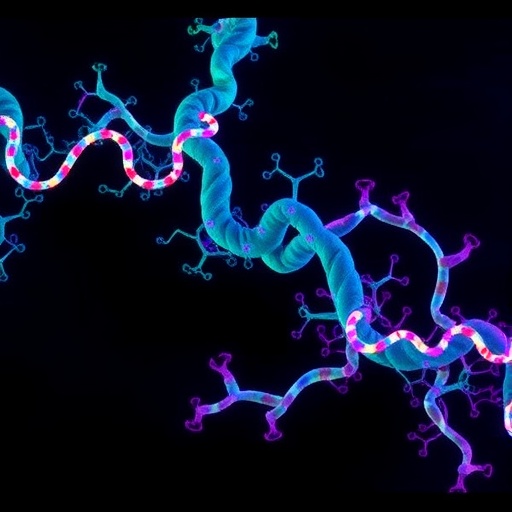
Traditional fluorescent nanobodies, despite their specificity, suffer from a ubiquitous problem: they emit fluorescence regardless of their binding state. This indiscriminate glow creates a diffuse background, obscuring critical intracellular details and limiting the resolution of protein localization and dynamics studies. To circumvent this, the team engineered a novel class of probes termed VIS-Fbs (visible-spectrum target-stabilizable fluorescent nanobodies). These innovative molecules are designed to degrade swiftly unless stabilized through binding to their specific protein targets. This degradation-driven selectivity ensures that only the bound nanobodies remain fluorescent, dramatically slashing background signal levels by up to 100 times and yielding unprecedented sharpness in live imaging.
The versatility of the VIS-Fb system is remarkable. The researchers crafted probes that span nearly the entire visible light spectrum—from blue to far-red—thereby enabling simultaneous multicolor tracking of multiple proteins or cellular activities within the same living cell. This spectral range offers an expansive toolkit for dissecting complex biological processes with unparalleled spatial resolution. Such multiplexing capacity is crucial for unraveling the interactions and co-localization of proteins within subcellular compartments, a feat previously hampered by probe limitations.
Beyond spectral diversity, the VIS-Fb platform is modular and adaptable. By integrating over twenty distinct fluorescent proteins and biosensors across various nanobody scaffolds, the scientists established a robust engineering framework to customize probes for diverse experimental scenarios. This modularity allows researchers to tailor the probes for different protein targets, cellular environments, and functional readouts. Notably, some VIS-Fb variants are engineered for optogenetic control, permitting researchers to switch fluorescence on or off with light stimulation. This capacity not only enables high-resolution temporal tracking of protein behavior but also facilitates dynamic perturbation studies in live systems.
Adding to its functional repertoire, the system incorporates biosensors sensitive to ions and metabolites, translating protein localization events into real-time reports of cellular activity. This dual capability allows for simultaneous visualization of protein presence and functional state, offering a holistic picture of intracellular signaling and metabolic flux. Furthermore, the combination of stable reference signals with these activity-sensitive elements enables ratiometric measurements, enhancing quantification accuracy in complex biological milieus, including densely packed brain tissues.
The practical applications of VIS-Fbs were validated in diverse live models. In mammalian neurons and astrocytes, the nanobody probes delivered high-fidelity imaging of central nervous system activity during behavioral assays, highlighting their utility for neuroscience research. In zebrafish embryos, these probes illuminated developmental dynamics and responses to pharmacological agents targeting cell signaling pathways, showcasing their versatility across species and research domains.
This innovation addresses a critical bottleneck in cellular imaging technology by reconciling the need for both specificity and clarity. By structurally linking nanobody stability to target engagement, the VIS-Fb system elegantly ensures that fluorescence signals represent authentic biological interactions rather than background artifacts. This specificity is pivotal for studying transient or low-abundance proteins that often evade detection with conventional methods.
According to Vladislav Verkhusha, Ph.D., co-corresponding author and professor of genetics at Einstein, the elimination of background fluorescence “opens the door to studying complex biological processes, such as cell signaling, development, and disease progression, in new ways.” His sentiments underscore the broader impact of VIS-Fbs—not simply as an imaging tool, but as a gateway to deeper mechanistic insights into cellular function and pathology.
The design strategy behind VIS-Fbs exemplifies a convergence of protein engineering, fluorescence biochemistry, and live-cell imaging technology. By harnessing the innate specificity of nanobodies and fusing it with a degradable scaffold linked to target binding, the researchers have created a self-regulating fluorophore. This concept could be extended to numerous molecular systems where minimizing background fluorescence is essential, such as in the visualization of intracellular pathogens, protein aggregates, or signaling microdomains.
The implications for biomedical research are profound. High-resolution, low-noise imaging enables more accurate mapping of protein interactions, post-translational modifications, and dynamic cellular responses. This refined resolution is particularly critical in neurobiology, immunology, and developmental biology, where subtle spatial and temporal changes underpin function and disease. Moreover, the VIS-Fb platform’s adaptability suggests future extensions into clinical diagnostic imaging or targeted therapeutic monitoring.
This work not only delivers a transformative imaging methodology but also establishes a paradigm for future probe development. The integrative approach combining target-induced stability, spectral multiplexing, optogenetic control, and biosensing sets a new benchmark for precision and functionality in live-cell imaging tools. As technology advances, such sophisticated molecular tools will increasingly bridge the gap between static molecular snapshots and dynamic, high-resolution portraits of living biology.
Collaborators on this seminal study included Juliana Mendoça-Gomes, Ph.D., and Sofia de Oliveira, Ph.D. from Einstein; Erin Carey from the Salk Institute; and Olena Oliinyk, Ph.D., from the University of Helsinki. The research was generously supported by several funding agencies, including the National Institutes of Health, the Jane and Aatos Erkko Foundation, the Research Council of Finland, the Chan Zuckerberg Initiative Foundation, the NOMIS Foundation Neuroimmunology Initiative, and the Edwards-Yeckel Research Foundation.
In sum, the engineered fluorescent nanobody probes developed by this collaborative effort herald a new era of intracellular imaging, granting scientists a clearer, more precise window into the molecular choreography of life. This advancement promises to accelerate discovery across multiple fields by unveiling the spatial and temporal complexity of protein function within native cellular contexts.
Subject of Research: Cells
Article Title: “Synthetic multicolor antigen-stabilizable nanobody platform for intersectional labelling and functional imaging.”
News Publication Date: 22-Apr-2026
Web References: http://dx.doi.org/10.1038/s41592-026-03056-3
References: Published in Nature Methods
Image Credits: Albert Einstein College of Medicine
Keywords: Fluorescent proteins, Live cell imaging, Biosensors
Tags: advanced biomedical imaging toolsantibody-derived fluorescent probesengineered fluorescent probes for protein visualizationfluorescent nanobodies for live cell imaginghigh-resolution intracellular protein imaginglive tissue protein dynamicsmolecular biology imaging techniqueson-demand activation fluorescent probesprotein localization in living cellsreducing background noise in fluorescence imagingselective fluorescent probe degradationVIS-Fbs probe technology