Ultrasound imaging, a cornerstone of modern medical diagnostics, has long been recognized for its capability to visualize anatomical structures and monitor physiological processes in real time. Commonly employed in contexts ranging from prenatal monitoring to internal examinations, the conventional applications of ultrasound have traditionally been limited to relatively large-scale body structures. However, a revolutionary advancement has recently emerged from a collaborative research effort involving TU Delft, the Netherlands Institute for Neuroscience, and Caltech. This innovative work introduces a trailblazing technique capable of imaging living cells in three-dimensional detail, surpassing the constraints imposed by traditional imaging modalities.
The research team accomplished something that was once thought nearly impossible: they successfully employed ultrasound to image specifically labelled living cells within entire organs, achieving depth resolutions previously unattainable in standard ultrasound practices. By engineering a method known as nonlinear sound sheet microscopy, researchers have unlocked the potential to visualize biological phenomena at a micro-scale, which adds a new dimension to our understanding of cellular processes. Unlike existing imaging techniques, which often necessitate the removal and processing of samples—hence disrupting the natural activity of cells—this innovative ultrasound approach allows for real-time monitoring while maintaining the integrity of living tissues.
At the core of this novel imaging capability is the use of specialized sound-reflecting probes tech, a development facilitated by the Shapiro Lab at Caltech. These sophisticated probes, which are nanoscale gas-filled vesicles, serve to enhance the visibility of cellular structures when subjected to ultrasound waves. This is achieved thanks to a shell of engineered proteins—designed to optimize their brightness in images—allowing researchers to track and visualize living cells effectively. The significance of this advancement cannot be overstated, as it facilitates the examination of cancer cells in their natural environments.
A noteworthy feature of this technique is its ability to penetrate deep within opaque mammalian tissues. Traditional light-based imaging methods, such as light sheet microscopy, come with significant limitations, particularly when dealing with thick or opaque specimens. These methods can only capture imaging over superficial layers, often failing to penetrate deeper than 1 mm. In stark contrast, the nonlinear sound sheet microscopy method can glean insights from several centimeters beneath the surface of tissues, thereby enabling comprehensive non-invasive investigations of entire organs over prolonged periods.
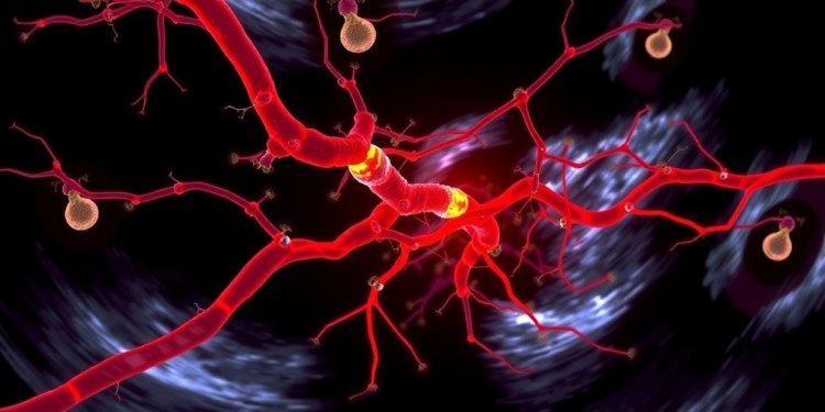
The implications of this technology extend beyond mere academic curiosity; they hold tremendous promise for practical medical applications. A pivotal application already identified by the research team is in the field of brain imaging, where they managed to utilize ultrasound alongside microbubble probes to observe capillaries in live brain tissues. This represents a groundbreaking advancement, laying the foundation for diagnosing conditions related to small vessel diseases, which are often difficult to assess using traditional imaging techniques. Given that microbubble probes have already received approval for human use, this ultrasound technique could soon transition from the laboratory to clinical settings, transforming the landscape of medical diagnostics.
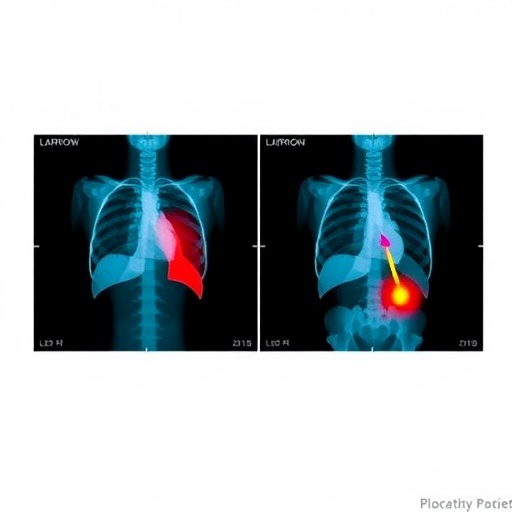
Furthermore, nonlinear sound sheet microscopy demonstrates potential for significant contributions to cancer research. The ability to distinguish between benign and malignant tissues can greatly enhance diagnostic precision and treatment efficacy. By revealing the necrotic cores of tumors, where cells perish due to a deficient oxygen supply, this imaging approach not only elucidates the current state of a tumor but can also assist medical professionals in evaluating the effectiveness of ongoing therapeutic interventions. This represents a critical advance in oncological treatment monitoring, moving beyond mere observation to actionable insight.
The intersection of ultrasound technology with micro-scale imaging insinuates a paradigm shift within both clinical and research domains. It empowers scientists and physicians to observe biological processes in real time, providing unique opportunities for longitudinal studies that were previously unimaginable. Researchers envisage that this method will offer vital data not just concerning cellular behavior in isolated environments, but under physiological conditions that closely resemble the body’s natural state, leading to more reliable and applicable findings.
Moreover, this technique promises to uncover previously hidden cellular dynamics critical to understanding complex illnesses. In conditions such as cancer, where cellular behavior and microenvironmental interactions play central roles in tumor progression and treatment outcomes, the capacity to visualize and analyze living cells in situ could yield breakthroughs in therapeutic development and application. Longitudinal analyses of cellular responses to various treatments will serve to optimize strategies tailored to individual patient needs and clinical contexts.
In summary, what we are witnessing with the development of nonlinear sound sheet microscopy is not merely an enhancement of existing technologies but rather a significant leap toward a more profound understanding of life at its core. As the research team continues to explore the myriad applications of this new method, the medical community can eagerly anticipate how this innovation will redefine diagnostics and treatment approaches across a range of health challenges.
This collaborative work exemplifies the power of interdisciplinary research, merging acoustics and biology to create a tool that delivers on the promise of enhanced understanding and the ability to intervene more effectively in health crises. As researchers refine these techniques and further validate their effectiveness in clinical settings, nonlinear sound sheet microscopy may redefine how we visualize and comprehend the intricate realities of living tissues.
The pathway forward will likely involve exploring the full capabilities of this imaging technique and expanding its applications into other areas of research and medicine. By bridging the gap between large-scale imaging and cellular-level insights, nonlinear sound sheet microscopy is on track to become an integral part of the toolkit for medical professionals and researchers alike, unveiling the mysteries of life itself with unprecedented clarity.
Subject of Research: Cells
Article Title: Revealing capillaries and cells in living organs with ultrasound
News Publication Date: 3-Apr-2025
Web References: Science
References: 10.1126/science.ads1325
Image Credits: Maayan Harel, Maresca Lab
Keywords: Ultrasound, 3D Imaging, Nonlinear Sound Sheet Microscopy, Living Cells, Cancer Research, Brain Imaging, Medical Diagnostics, Cellular Behavior, Oncological Treatment, Microbubbles, Neuroscience, Acoustic Probes.
Tags: advancements in medical diagnosticsCaltech biomedical innovationscollaborative research in medical imagingliving organ imaging techniquesnonlinear sound sheet microscopypreserving cellular integrity in imagingreal-time cellular monitoringrevolutionary ultrasound applicationsthree-dimensional imaging of living cellsTU Delft neuroscience researchultrasound imaging technologyvisualization of micro-scale biological phenomena

