
Researchers at the Weizmann Institute of Science have announced a groundbreaking advancement in the field of biomedical imaging, introducing an innovative AI-assisted technology known as CombPlex. This cutting-edge technique enables the simultaneous visualization and quantification of a significantly heightened number of proteins within individual cells in tissue samples. Published in the esteemed journal Nature Biotechnology, this research could revolutionize our understanding of cellular composition in various tissues, particularly in the context of disease.
The scientific need for comprehensive protein measurement is underscored by Dr. Leeat Keren, who led the research team from the Molecular Cell Biology Department at the Weizmann Institute. The ability to measure numerous proteins simultaneously is crucial for understanding the functioning of different tissues. By analyzing the interactions and communications among diverse cell types within a tissue sample, researchers can gain insights into the intricacies of disease processes. For instance, in cancerous tissues, the cellular ecosystem consists not only of tumor cells but also healthy cells and immune cells. Understanding these interactions is vital for tailoring effective therapies and identifying patient prognoses.
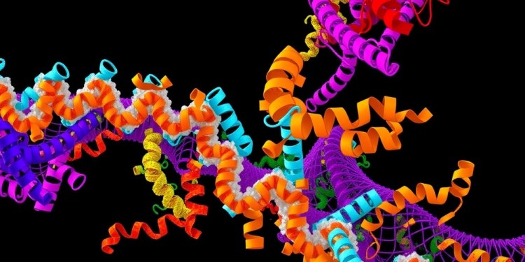
CombPlex empowers scientists to delve deeper into the cellular makeup of tissues than traditional methods, which typically only allow for the analysis of three or four proteins at a time. The new technology reveals the potential to image up to hundreds of proteins concurrently, vastly expanding the scope of biomedical research. Importantly, CombPlex does not require additional instrumentation, making it a highly accessible tool for researchers and clinicians alike. This accessibility could lead to broader adoption and application in various medical and research settings.
The development of CombPlex emerged from the limitations posed by traditional imaging techniques. Conventional methods, which process fluorescent probes for protein tagging, face significant challenges when attempting to visualize multiple proteins within the same sample. The overlapping colors of different probes can obscure critical details, complicating the interpretation of results. While cyclic fluorescence techniques allow researchers to iteratively image proteins, these approaches are time-consuming and often lead to diminishing returns as only a limited number of proteins can be analyzed ultimately.
Dr. Keren’s vision for enhancing imaging techniques involved overcoming these limitations by capturing a more comprehensive picture of tissue samples. Drawing an analogy between imaging proteins and photographing various objects in a room, Dr. Keren envisioned a single snapshot that encapsulates the entirety of protein interactions rather than a fragmented collection of images. This fundamental shift in approach paved the way for the CombPlex technology, which aims to facilitate the analysis of numerous proteins simultaneously.
To achieve this ambitious goal, the research team employed a combinatorial approach to protein labeling. This method involves attaching multiple fluorescent tags to each protein, thus generating unique combinations—akin to barcodes—that allow for greater resolution in imaging. By leveraging the power of combination and permutation, the researchers could vastly increase the number of distinct proteins analyzed with a limited array of fluorescent colors.
However, a new challenge emerged: the complexity of viewing overlapping barcodes created from the combinatorial labeling. Keren and her team recognized that artificial intelligence could hold the key to deciphering the entangled signals produced during imaging. By training an AI algorithm to learn the expression patterns of different proteins within tissue images, the researchers could resolve overlapping signals, effectively isolating individual protein images that had previously appeared as insurmountably complex.
In collaboration with Dr. Shai Bagon of the Weizmann Center for Artificial Intelligence, the team designed an experimental methodology complemented by a robust AI algorithm. The researchers trained a deep neural network using simulated data derived from various fluorescent protein images. This deep learning approach enabled the model to distinguish and unpack the intertwined visual signals, providing a clearer and more accurate depiction of protein presence within tissue samples.
The resultant technology, CombPlex, yields impressive results as it accurately quantifies multiple proteins at the cellular level. Its capability to transform seemingly chaotic fluorescent tangle images into distinct individual protein depictions marks a significant milestone for biomedical research. As CombPlex integrates seamlessly with conventional fluorescent microscopes, it promises to enhance the efficiency and depth of protein analysis in both laboratory and clinical environments.
The advantages of the CombPlex method extend beyond sheer imaging capability. It offers a faster alternative to obtaining comprehensive protein data, potentially reducing the time required for detailed analysis from weeks to just a couple of days. This efficiency could dramatically accelerate the pace of research and contribute to timely clinical decision-making. As clinicians increasingly seek precision in diagnostics and treatments, CombPlex stands out as a transformative tool that could redefine standard practices in pathology.
The theoretical framework supporting CombPlex suggests that researchers can capture up to 2^n – 1 proteins using merely n tags, exemplifying the exponential potential of this technology. For instance, employing three tags unlocks the possibility of analyzing up to seven proteins, while five tags enable the analysis of 31 proteins. Although real-world applications may encounter various challenges, Keren’s research team successfully demonstrated the effective measurement of 22 proteins using combinations of five distinct tags.
Guided by the Weizmann Institute’s translational research unit, Bina, the team navigated the complexities of developing an applicative technology like CombPlex. The excitement and enthusiasm from field experts affirmed the potential of CombPlex, further motivating the research team. As Dr. Sharon Fireman, head of Bina, noted, the positive response from experts highlights the significance of this groundbreaking advancement.
With a talented multidisciplinary team comprised of students and faculty from diverse scientific backgrounds, including biochemistry, bioinformatics, and mathematics, CombPlex embodies a collaborative effort towards scientific progress. Their collective dedication to unraveling the complexities of protein interactions illustrates the potential for interdisciplinary approaches to yield transformational outcomes in contemporary biomedical research.
As CombPlex heralds a new era in tissue analysis, the broader implications of such advancements cannot be overstated. As researchers continue to unlock the intricacies of cellular interactions and reframe our understanding of diseases, the advent of AI-assisted imaging technologies like CombPlex will undoubtedly catalyze further innovations in personalized medicine, therapeutic development, and diagnostic precision. With its promise of comprehensive and efficient analysis, CombPlex represents a beacon of hope for researchers and clinicians aiming to enhance patient outcomes through improved data visualization and interpretation.
As the scientific community embraces the transformative potential of technologies like CombPlex, it becomes evident that the future of biomedical research lies at the intersection of artificial intelligence and innovative imaging techniques. This harmonious convergence not only elevates our understanding of biological processes but also paves the way for a more nuanced approach to medicine, where data-driven insights will inform clinical care and lead to breakthroughs in disease treatment and prevention.
In conclusion, the introduction of CombPlex is more than just a technical advancement; it embodies a paradigm shift in the way we study and interpret the intricate dynamics of cellular interactions within tissues. The potential to visualize and quantify an unprecedented number of proteins holds immense implications for both basic research and clinical applications alike. As researchers delve deeper into the cellular landscape, they will be better equipped to uncover the complexities of health and disease, ultimately advancing the frontiers of biomedical science into uncharted territories of knowledge and understanding.
Subject of Research: Artificial Intelligence in Biomedical Imaging
Article Title: High-dimensional imaging using combinatorial channel multiplexing and deep learning
News Publication Date: 25-Mar-2025
Web References: Nature Biotechnology
References: 10.1038/s41587-025-02585-0
Image Credits: Weizmann Institute of Science
Keywords: AI, biomedical imaging, protein quantification, fluorescent microscopy, cellular analysis, combinatorial labeling, deep learning, tissue composition, cancer research, personalization, clinical applications.
Tags: advancements in tissue sample analysisAI-assisted biomedical imagingCombPlex technology for protein visualizationcomprehensive protein measurement methodsenhancing disease prognosis through protein analysisimplications for tailored cancer therapiesinnovative techniques in molecular cell biologyinsights into cancer tissue interactionsNature Biotechnology publication on protein researchsimultaneous protein quantification in cellsunderstanding cellular composition in diseasesWeizmann Institute of Science research


