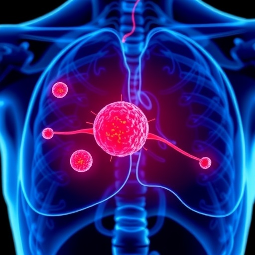
A groundbreaking study from the University of Cambridge is shedding new light on the earliest events that determine whether a microscopic tumour survives or succumbs in its nascent stages. By investigating the interplay between emerging tumour cells and the healthy cells in the surrounding supportive tissue, scientists are unraveling the complex microenvironmental dynamics that influence cancer initiation and progression. This pioneering work offers profound insights that could revolutionize early cancer diagnosis and prevention strategies, particularly for oesophageal cancer, a malignancy often detected too late for effective intervention.
The genesis of cancer has long been understood as a consequence of genetic mutations within cells that lead to unregulated growth and evasion of programmed cell death. However, the mere presence of these mutations in cells does not inexorably lead to full-blown malignancy, as similar mutations accumulate harmlessly in some healthy individuals, especially with age. The critical question tackled by the Cambridge team was: what additional biological factors govern whether mutated cells persist, proliferate, or are eliminated by natural tissue defenses?
Drilling down to the very earliest moments when a tumour first appears, the researchers focused on the esophageal epithelium in a mouse model. By exposing mice to a tobacco-derived carcinogen—a principal risk factor for human oesophageal cancer—they induced genetic alterations in the cells lining the oesophagus, triggering the formation of microscopic tumours comprising just a handful of mutant cells. Most of these nascent cellular clusters disappeared spontaneously, mirroring clinical observations of subclinical tumours in humans that never develop into cancers.
Employing state-of-the-art methodologies, including high-resolution confocal microscopy, single-cell RNA sequencing, and sophisticated genetic lineage tracing, the team meticulously tracked the fate of these early tumours over time. They also honed three-dimensional tissue culture systems that replicated the tumour microenvironment, enabling them to dissect molecular exchanges between the tumour cells and neighboring stromal cells. What emerged from this detailed cellular portrait was a revealing narrative of bidirectional communication that challenges traditional mutation-centric cancer paradigms.
Central to their discovery was the role of fibroblasts—resident “first-responder” cells in the underlying connective tissue traditionally known for their function in wound repair and extracellular matrix deposition. The researchers observed that the tiny tumour clusters emit distress signals that activate these fibroblasts. Upon activation, the fibroblasts undergo phenotypic changes akin to those seen in wound healing, generating a fibrotic scaffold composed of extracellular matrix proteins tightly enveloping the tumour cells. This fibrotic niche effectively functions as a protective cocoon, shielding the developing tumour from immune clearance and fostering its persistence.
Intriguingly, this remodeled microenvironment does not merely shelter mutated cells; it appears capable of inducing tumour-like characteristics in otherwise genetically normal epithelial cells. This phenomenon underscores an emerging concept that cancer development is not dictated solely by cell-intrinsic genetic aberrations but is profoundly shaped by the cellular and molecular makeup of the surrounding tissue landscape. Such pre-cancerous niches can foster cellular behaviors that predispose tissues to malignancy, long before overt genetic transformations accumulate.
Crucially, the team demonstrated the translational relevance of these findings by examining human oesophageal cancer specimens obtained at early stages. Mirroring the murine data, these human tissues exhibited clusters of stressed tumour cells surrounded by fibrotic scaffolds, affirming that the identified tissue remodeling mechanism operates in human disease as well. This cross-species concordance reinforces the potential for these insights to inform human clinical strategies.
The significance of the pre-cancerous niche was further established through functional experiments where the investigators disrupted the communication pathways between tumour cells and fibroblasts. When the distress signaling was pharmacologically blocked, the formation of the fibrotic scaffold was markedly impaired, and the survival rate of early tumours plummeted. This finding highlights the therapeutic potential of targeting tumour-stroma crosstalk in cancer prevention, a conceptual shift from traditional approaches focused exclusively on mutated cancer cells.
Beyond therapeutic implications, this research opens new avenues for early cancer detection. The team identified candidate biomarkers—essentially molecular “red flags” generated by both tumour and stromal cells during niche formation—that may enable clinicians to diagnose oesophageal cancer at a stage when it is far more amenable to treatment. Early-stage detection is particularly crucial for oesophageal cancer, given its typically poor prognosis when diagnosed late.
The study also disrupts conventional wisdom that frames early tumour survival as predominantly determined by the mutant cells’ own oncogenic properties. Instead, it positions the healthy tissue’s response as a critical determinant of whether a tumour is eradicated or nurtured into a clinically significant cancer. This paradigm shift underscores the importance of viewing cancer initiation through the lens of tissue ecology and intercellular communication networks.
From a biological perspective, the findings demonstrate how the healing and regenerative processes of normal tissue can be hijacked by emerging cancerous cells to create a microenvironment conducive to tumour progression. The parallels drawn between wound healing and tumour niche formation reveal the dual-edged nature of cellular response mechanisms, capable of either restoring tissue integrity or inadvertently facilitating disease.
This research was enabled by an interdisciplinary collaboration among stem cell biologists, developmental physiologists, and cancer researchers at the Cambridge Stem Cell Institute and the Department of Physiology, Development and Neuroscience. The experimental rigor and innovative use of both in vivo and in vitro models provide a robust framework for further exploration of the early tumour microenvironment and its role in cancer biology.
Looking forward, further work is needed to identify the precise molecular mediators of the distress signals and fibroblast activation. Pinpointing these pathways could yield novel drug targets for early intervention, potentially transforming cancer prevention strategies. Additionally, clinical studies to validate the proposed biomarkers in human populations will be pivotal for translating this work into improved diagnostic tools.
Funded by Worldwide Cancer Research, the Wellcome Trust, the Royal Society, the Medical Research Council, and the Isaac Newton Trust, this study exemplifies how foundational research can inform future clinical breakthroughs. The findings align with broader ambitions at the University of Cambridge, including efforts to establish a dedicated Cancer Research Hospital focused on early detection and precision treatment.
In sum, this landmark study reshapes our understanding of how cancers take root, emphasizing that it is not solely the rogue genetic mutations but the collaborative behavior of mutated cells and their healthy neighbors that dictates cancer’s fate. Unraveling these early dialogues between tumour and tissue may be humanity’s best chance to interrupt cancer before it fully takes hold.
Subject of Research: Animals
Article Title: Precancerous niche remodelling dictates nascent tumour persistence
News Publication Date: 4-Mar-2026
Web References: https://doi.org/10.1038/s41586-026-10157-8
References: Skrupskelyte, G et al. Precancerous niche remodelling dictates nascent tumour persistence. Nature; 4 March 2026; DOI: 10.1038/s41586-026-10157-8
Keywords: Cancer, tumour microenvironment, oesophageal cancer, fibroblasts, early detection, tumour persistence, fibrosis, cancer prevention, tissue remodeling, single-cell RNA sequencing
Tags: cancer prevention strategies in esophageal cancerearliest stages of tumor survivalearly cancer detection in esophageal cancerearly cancer diagnosis innovationsesophageal epithelium mouse model studygenetic mutations and cancer progressioninterplay between tumor cells and healthy tissuemolecular mechanisms of tumor persistencenatural tissue defenses against tumorspioneering cancer research from University of Cambridgetobacco carcinogen impact on cancertumor microenvironment and cancer initiation
