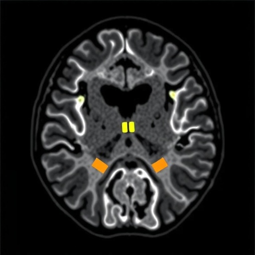
Sturge-Weber Syndrome (SWS) is a complex neurocutaneous disorder characterized by a unique triad of neurological and cutaneous manifestations. One of the most defining features of SWS is the presence of a facial capillary malformation, often referred to as a port-wine stain, which typically affects the forehead and upper eyelid on the side of the brain where the abnormal vascular development occurs. Beyond its outward appearance, SWS has profound implications for neurological development due to the underlying vascular anomalies and associated hypoxia, making its early diagnosis imperative for timely intervention.
In the pursuit of understanding SWS, researchers have put significant effort into elucidating the magnetic resonance imaging (MRI) features associated with the syndrome. MRI serves as an invaluable tool in identifying both the structural and functional anomalies present in affected individuals. Recent studies, particularly the comprehensive review conducted by Cerron-Vela et al., aim to catalogue these imaging findings, highlighting the evolution of vascular changes from infancy through adulthood, and drawing correlations between imaging findings and clinical outcomes.
On MRI, patients with SWS typically display leptomeningeal angiomas, which are abnormal growths of blood vessels in the protective membranes surrounding the brain. These angiomas can lead to localized ischemia and other complications, significantly impacting the patient’s neurological health. Over time, these vascular changes may progress, culminating in cortical atrophy, white matter changes, and even the development of tissue necrosis, which can further exacerbate clinical symptoms and complicate treatment plans.
One of the most critical aspects of the research is the focus on the timing and nature of vascular responses to the underlying pathology. Early vascular responses may include hyperperfusion, where affected areas of the brain exhibit increased blood flow. This finding can sometimes mask underlying ischemic issues during initial imaging studies. As the condition progresses, however, hypoperfusion tends to dominate, posing challenges for patient management and necessitating careful monitoring of neurological status.
Equally pivotal in the exploration of SWS is the observation of neurological sequelae. Children with Sturge-Weber Syndrome may encounter various issues ranging from developmental delays and seizures to higher risks of stroke or other cerebrovascular accidents. The relationship between the severity of MRI findings and the extent of these neurological complications has sharpened diagnostic accuracy and paved the way for better clinical outcomes through targeted therapies.
As imaging technology advances, the ability to detect subtle changes in brain architecture becomes increasingly sophisticated. Advanced techniques such as diffusion tensor imaging (DTI) are now being employed to assess microstructural brain changes that are not visible on conventional MRI. DTI can provide insights into the integrity of white matter tracts that could be compromised in individuals with SWS. This information not only aids in diagnosis but also informs treatment strategies tailored to individual patient needs.
Another intriguing area of study delves into the genetic and environmental factors that contribute to the expression and severity of Sturge-Weber Syndrome. Until recently, the underlying etiology was poorly understood, which often left patients and families in uncertainty. Genetic studies are now revealing key mutations associated with SWS, opening avenues for innovative therapies that could address the root cause of the disorder rather than solely managing symptoms.
The importance of interdisciplinary care cannot be overstated in the management of SWS. Neurologists, dermatologists, radiologists, and pediatricians must work collaboratively to form a cohesive care plan that optimizes outcomes for these patients. The integration of insights gleaned from MRI studies with comprehensive clinical evaluations helps to tailor individualized management strategies that take into account the multifaceted nature of the syndrome.
In investigating the therapeutic landscape, researchers are increasingly exploring options for intervention that may alter the course of the disease. Early intervention strategies, employing techniques from pharmacological treatments to surgical options for resecting problematic vascular channels, have shown promise in enhancing quality of life and reducing the impact of neurological sequelae. Continuous follow-ups with neuroimaging allow for monitoring of disease progression and treatment efficacy.
Furthermore, the role of patient and family education in navigating the complexities of Sturge-Weber Syndrome cannot be overlooked. Comprehensive educational resources are essential for families to understand the implications of the diagnosis, resulting in better adherence to treatment plans and proactive measures that may prevent complications. As awareness breeds understanding, both patients and families can become vital advocates for their healthcare needs.
Notably, the strides being made in the understanding of Sturge-Weber Syndrome through imaging studies are contributing to an expanded knowledge base. The lessons learned from one congenital vascular disorder can potentially be translated to other similar conditions, fostering a broader understanding of vascular anomalies and their implications across patient populations. This ripple effect of knowledge dissemination serves as a foundation for future research endeavors.
The socio-emotional impact of living with Sturge-Weber Syndrome is another vital piece of the puzzle. Patients frequently navigate challenges not only related to their physiological symptoms but also psychological hurdles, such as fear of stigma or the unpredictability of their condition. Therefore, addressing the psychosocial aspects of care is as crucial as medical management, ensuring comprehensive support structures are in place to encourage thriving lifestyles.
In conclusion, Cerron-Vela et al.’s review of MR imaging features in Sturge-Weber Syndrome provides an essential resource for practitioners involved in the management of this complex disorder. Its implications resonate beyond the confines of radiology, intertwining with genetics, clinical practice, and psychosocial support, creating a mosaic that demands a holistic approach. As our understanding of SWS deepens, we remain poised to transform the landscape of patient care, ultimately leading to enhanced outcomes for those affected by Sturge-Weber Syndrome.
Subject of Research: Sturge-Weber Syndrome and its MRI features
Article Title: Beyond the leptomeningeal angioma: a comprehensive review of MR imaging features of Sturge-Weber Syndrome, from early vascular responses to tissue necrosis.
Article References:
Cerron-Vela, C.R., Manteghinejad, A. & Andronikou, S. Beyond the leptomeningeal angioma: a comprehensive review of MR imaging features of Sturge-Weber Syndrome, from early vascular responses to tissue necrosis.
Pediatr Radiol (2025). https://doi.org/10.1007/s00247-025-06402-3
Image Credits: AI Generated
DOI: https://doi.org/10.1007/s00247-025-06402-3
Keywords: Sturge-Weber Syndrome, MRI, leptomeningeal angioma, neurocutaneous disorder, neuroimaging
Tags: clinical outcomes in Sturge-Weber Syndromeearly diagnosis of Sturge-Weber Syndromefacial capillary malformationhypoxia and SWSleptomeningeal angiomasMRI imaging featuresneurocutaneous disordersneurological development implicationsport-wine stainstructural and functional anomaliesSturge-Weber Syndromevascular anomalies in SWS


