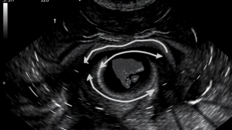
Ovarian endometrioma, one of the most stubborn and clinically consequential manifestations of endometriosis, has long forced gynecologists into an uncomfortable trade-off. Surgery can remove the chocolate-colored cysts that plague millions of women worldwide, but every pass of the scalpel near ovarian tissue risks chipping away at the very reserve of eggs that patients, particularly those facing infertility, desperately want to protect. Now a comprehensive review published in the Journal of Ovarian Research argues that the answer to this dilemma may lie not in a new drug or a new surgical instrument, but in something far more familiar: the ultrasound probe. A team of researchers from the Sichuan Clinical Research Center for Cancer in Chengdu, China, led by Yi Zhu and colleagues, has mapped out how transvaginal ultrasound could serve as the backbone of decision-making for ultrasound-guided sclerotherapy, a minimally invasive alternative that drains the cyst and scars its lining shut without cutting into the ovary at all.
The procedure itself is elegantly simple in concept. Under real-time ultrasound guidance, a needle is passed, usually transvaginally, into the endometrioma, the thick dark fluid is aspirated, and a sclerosing agent, most commonly absolute ethanol, is instilled into the cavity. The ethanol destroys the endometrial lining that secretes the cyst’s contents, effectively gluing the walls together and dramatically lowering the chance that the lesion refills. Because the ovarian cortex and its follicles are never incised, the technique has attracted growing interest among fertility specialists who worry that repeated cystectomies can measurably deplete anti-Müllerian hormone levels and antral follicle counts, the two laboratory and imaging markers of ovarian reserve. Yet sclerotherapy has never achieved the clinical standing of laparoscopic surgery, and the new review makes clear why: the evidence base is fragmented, with studies differing wildly in who gets treated, how the procedure is performed, how much sclerosant is used, and how success and recurrence are even defined.
What the Chinese team proposes is a standardized framework in which ultrasound is not merely a guidance tool for the moment of needle insertion but an active decision-making partner across the entire care pathway. The review, which synthesized English-language literature up to May 2026 from PubMed, Embase, Web of Science, and Scopus, organizes the ultrasound contribution into distinct phases: diagnosis, risk stratification, patient selection, procedural planning, treatment guidance, and post-treatment surveillance. This staging matters because each phase demands different information from the same imaging modality, and because errors made early in the pathway, such as mistaking a malignant tumor for an endometrioma, can be catastrophic if a clinician proceeds to puncture and sclerose the wrong lesion.
Diagnosis is where transvaginal ultrasound already reigns supreme. The characteristic appearance of an endometrioma, a well-defined cyst with homogeneous low-level internal echoes, so-called ground-glass echogenicity, and no internal vascular flow, is recognizable to any trained sonographer. But the review emphasizes that the real diagnostic task is exclusion. Endometriomas have a notorious gallery of mimickers, including hemorrhagic corpus luteum cysts, dermoid cysts, fibromas, and, most dangerously, malignant ovarian tumors that can present with atypical cystic features. Structured approaches such as the International Ovarian Tumor Analysis simple rules and the IOTA ADNEX model allow clinicians to classify adnexal masses with high accuracy and to flag red features, solid components, papillary projections, ascites, or increased color Doppler flow, that mandate referral for magnetic resonance imaging or oncological assessment rather than any intervention. Only after malignancy is excluded can the conversation about sclerotherapy responsibly begin.
Once the diagnosis is secure, ultrasound shifts to a second, less celebrated role: risk stratification and patient selection. Here the review draws on the International Deep Endometriosis Analysis framework, which uses ultrasound to map not just the cyst itself but the entire pelvic environment. Is the ovary mobile or fixed to the uterosacral ligament? Is there deep endometriosis infiltrating the rectovaginal septum? Are there signs of hydrosalpinx, tubo-ovarian abscess, or dense adhesions that would make a transvaginal puncture hazardous or futile? These findings directly determine candidacy. An ideal candidate for sclerotherapy is typically a woman with a unilateral or bilateral endometrioma of sufficient size, a mobile ovary that can be safely reached by the needle, no suspicious features, and a clinical priority of preserving ovarian reserve, whether because she is pursuing assisted reproduction or because she has already lost ovarian function on the contralateral side. Conversely, ultrasound evidence of deep infiltrating disease, frozen pelvis, or an ovary inaccessible without traversing bowel or vessels steers the patient back toward laparoscopy or medical management.
The procedural planning phase is where the review’s technical depth becomes most apparent. Ultrasound provides the measurements that dictate the mechanics of the intervention: cyst diameter determines needle gauge and the volume of aspirated fluid, which in turn informs the volume and dwell time of the ethanol instillation. Most published protocols rinse the cavity with ethanol for a fixed number of minutes, on the logic that contact time between the sclerosant and the cyst lining governs the completeness of epithelial destruction. Too brief an exposure invites recurrence; too prolonged an exposure risks ethanol leaking past the needle tract and injuring surrounding tissue. Real-time imaging allows the operator to confirm that the needle tip remains intracystic throughout, that the cyst wall does not collapse prematurely, and that the sclerosant is fully recovered before withdrawal. Doppler imaging adds a further safety check, ensuring the puncture path avoids the ovarian hilar vessels and the iliac vasculature that lie in harm’s way during a transvaginal approach.
For women pursuing in vitro fertilization or intracytoplasmic sperm injection, the timing question is especially delicate. Aspiration and sclerotherapy performed too close to ovarian stimulation can theoretically impair the response of the treated ovary, while an untreated endometrioma can complicate egg retrieval by contaminating follicular fluid or becoming infected. The review suggests that ultrasound-based assessment of the lesion’s size, accessibility, and behavior over serial scans can help clinicians individualize the sequencing of sclerotherapy relative to assisted reproductive technology cycles, although it candidly acknowledges that the literature does not yet provide definitive answers on optimal timing. This honesty is a recurring theme: the authors repeatedly note that heterogeneity in patient selection criteria, sclerosant exposure protocols, response definitions, and follow-up intervals makes head-to-head comparison of published studies treacherous.
Surveillance after treatment is the final and arguably most underdeveloped link in the chain. Here ultrasound again carries the load, with serial transvaginal scans tracking whether the treated cyst remains collapsed, refills, or regrows. Recurrence definitions vary across studies, from any re-accumulation of fluid to symptomatic recurrence requiring re-intervention, and follow-up windows range from a few months to several years. The review argues that a standardized ultrasound surveillance schedule, combined with laboratory markers such as anti-Müllerian hormone to monitor whether ovarian reserve is genuinely preserved, would allow the field to finally generate comparable outcome data. It also points out that ultrasound can detect the rare but serious complications of the procedure, including pelvic infection, bleeding, and, in theory, iatrogenic endometriosis dissemination along the needle tract, early enough for effective management.
The bigger picture painted by the review is one of cautious optimism. Ultrasound-guided sclerotherapy appears feasible and attractive for selected women, particularly when preservation of ovarian reserve is the overriding priority, and it can be performed on an outpatient basis without general anesthesia, at a fraction of the cost of laparoscopic surgery. But the authors stop well short of declaring it ready to displace surgery. Instead, they issue a call to action: a standardized, ultrasound-anchored framework for candidate selection, procedural consistency, and post-treatment monitoring could transform sclerotherapy from a niche technique practiced unevenly across centers into a rigorously defined option within the endometriosis treatment arsenal. For the estimated ten percent of reproductive-age women living with endometriosis, many of whom face the double threat of pain and infertility, that framework could mean the difference between an ovary sacrificed and an ovary saved, decided not by a surgeon’s judgment alone but by the quiet, real-time evidence of a probe held against the pelvic wall.
Subject of Research: Ultrasound-guided decision-making for minimally invasive sclerotherapy of ovarian endometrioma
Article Title: Ultrasound-based decision-making for minimally invasive sclerotherapy of ovarian endometrioma
Article References: Zhu, Y., Mo, Y., Zhang, J., Yang, W., Li, Y., Liu, C., & Jia, S. (2026). Ultrasound-based decision-making for minimally invasive sclerotherapy of ovarian endometrioma. Journal of Ovarian Research. https://doi.org/10.1186/s13048-026-02257-w
Image Credits: AI Generated
DOI: 10.1186/s13048-026-02257-w
Keywords: ovarian endometrioma, endometriosis, transvaginal ultrasound, sclerotherapy, ethanol sclerotherapy, ovarian reserve, infertility, assisted reproduction, minimally invasive treatment, ovarian cysts, fertility preservation, gynecology
Cite Scienmag News
APA MLA Chicago
Ophelia Keating. (October 1, 2026). Ultrasound Takes Center Stage in Guiding Minimally Invasive Sclerotherapy for Ovarian Endometrioma. Scienmag. https://scienmag.com/ultrasound-takes-center-stage-in-guiding-minimally-invasive-sclerotherapy-for-ovarian-endometrioma/
Ophelia Keating. “Ultrasound Takes Center Stage in Guiding Minimally Invasive Sclerotherapy for Ovarian Endometrioma.” Scienmag, 1 October 2026, https://scienmag.com/ultrasound-takes-center-stage-in-guiding-minimally-invasive-sclerotherapy-for-ovarian-endometrioma/. Accessed 1 October 2026.
Ophelia Keating. “Ultrasound Takes Center Stage in Guiding Minimally Invasive Sclerotherapy for Ovarian Endometrioma.” Scienmag. October 1, 2026. https://scienmag.com/ultrasound-takes-center-stage-in-guiding-minimally-invasive-sclerotherapy-for-ovarian-endometrioma/
Copy citation Download RIS
Tags: advantages of ultrasound over traditional surgeryassisted reproductionendometriosisethanol sclerotherapyethanol sclerotherapy for ovarian cystsfertility preservationgynecologyinfertilityinfertility preservation in endometrioma treatmentminimally invasive treatmentminimally invasive treatment for endometriosisnon-surgical management of ovarian endometriomasovarian cyst drainage and sclerosisovarian cystsovarian endometriomaOvarian Reservepreserving ovarian reservereal-time ultrasound guidance in minimally invasive gynecologysclerotherapytransvaginal ultrasoundtransvaginal ultrasound in gynecological proceduresultrasound imaging in ovarian lesion managementultrasound-assisted ovarian cyst ablationultrasound-guided ovarian endometrioma sclerotherapy