In a groundbreaking study, researchers have made significant advancements in understanding abdominal compartment syndrome (ACS) following omphalocele repair, a common congenital condition affecting newborns. The findings, published in the journal Pediatrics Radiology, shed light on the critical role of ultrasound imaging in diagnosing this serious post-operative complication. This research underscores the importance of early detection and intervention in improving outcomes for affected infants.
Abdominal compartment syndrome is characterized by the increased pressure within the abdominal cavity, leading to diminished organ perfusion and function. This condition can arise as a result of various surgical interventions, most notably following repair procedures for omphalocele, which is a defect where abdominal organs protrude through the abdominal wall. Given the delicate nature of neonatal patients, timely diagnosis is imperative to prevent severe morbidity and mortality.
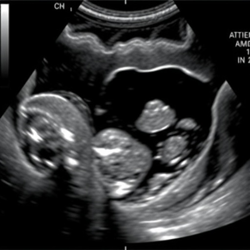
The researchers, led by de Souza Pires and colleagues, utilized ultrasound as a non-invasive imaging technique to assess infants post-omphalocele repair. This approach is particularly valuable in pediatrics, where minimizing invasive procedures is a priority. The study’s findings highlight how ultrasound can effectively visualize changes in the abdominal cavity that signify the onset of compartment syndrome.
In their research, the team established specific ultrasound criteria for identifying ACS, which could serve as a guideline for practitioners in the neonatal intensive care units. This is particularly crucial as neonates are often unable to verbalize their discomfort or distress, making traditional diagnostic methods less effective. The ability to rely on ultrasound can help clinicians make informed decisions about the management of these vulnerable patients.
Our understanding of abdominal compartment syndrome has evolved substantially over the years, yet the complexities involved in the post-operative care of neonates remain challenging. The new ultrasound guidelines proposed by this study could enhance clinical practices by providing a framework for monitoring patients who have undergone omphalocele repair. This could lead to earlier interventions and a reduction in the long-term complications associated with ACS.
Moreover, the implications of this research extend beyond just the immediate post-operative period. By enabling clinicians to detect signs of ACS early, the findings can help set a precedent for better long-term management of patients with congenital defects. Successful management of these complications can lead to improved overall developmental outcomes for children affected by omphalocele.
The potential for ultrasound as a long-term monitoring tool is a key takeaway from this research. Not only does it allow for real-time assessment of the abdominal cavity’s condition, but it also provides valuable data that can be utilized for ongoing research into best practices for neonate care. This aligns with the broader trends in pediatric medicine that favor enhanced monitoring techniques and improved patient outcomes through technology.
In a clinical context, the introduction of ultrasound as a standard diagnostic tool could also change the dynamics of team-based care in neonatal units. As healthcare teams become more aware of the specific ultrasound indicators for abdominal compartment syndrome, a more collaborative approach to patient management may emerge. This could foster a shared responsibility among healthcare providers and result in better decision-making for patient care.
As the field of pediatric radiology continues to advance, studies like this one play an essential role in bridging the gap between surgical intervention and radiological assessment. The emerging data suggest that effective communication between surgeons and radiologists could enhance post-operative care pathways, allowing for a more integrated approach to pediatric surgery.
Additionally, it is essential to highlight the study’s methodology, which included a robust sample of patients and thorough follow-up evaluations. Future studies are encouraged to replicate these findings across diverse medical settings to further validate the use of ultrasound in diagnosing abdominal compartment syndrome. Key clinical questions remain, including optimal follow-up intervals and the integration of ultrasound assessments into existing care protocols.
This important work brings to the forefront the challenges faced by neonates recovering from repair surgeries like omphalocele, emphasizing the critical need for vigilance and responsiveness in their care. Advances in imaging techniques not only enable healthcare professionals to diagnose conditions more accurately but also provide children with a better chance at a bright future.
In conclusion, the findings from this study have far-reaching implications for pediatric care, particularly for the management of abdominal compartment syndrome following omphalocele repair. As the medical community strives toward improving patient outcomes, the integration of advanced imaging technologies like ultrasound will undoubtedly play a pivotal role in shaping the future landscape of pediatric health care.
Subject of Research: Abdominal compartment syndrome after omphalocele repair.
Article Title: Ultrasound findings of abdominal compartment syndrome after omphalocele repair.
Article References:
de Souza Pires, P., Cortada Lluelles, R. & Arenos, J. Ultrasound findings of abdominal compartment syndrome after omphalocele repair.
Pediatr Radiol (2025). https://doi.org/10.1007/s00247-025-06468-z
Image Credits: AI Generated
DOI: 17 November 2025
Keywords: Abdominal compartment syndrome, omphalocele repair, ultrasound, pediatric radiology, neonatology.
Tags: abdominal cavity pressure assessmentabdominal compartment syndrome diagnosiscongenital condition managementcritical care in infantsearly detection of ACSneonatal health outcomesnon-invasive imaging techniquesomphalocele repair complicationspediatric radiology advancementssurgical interventions in newbornsultrasound criteria for ACSultrasound imaging in pediatrics


