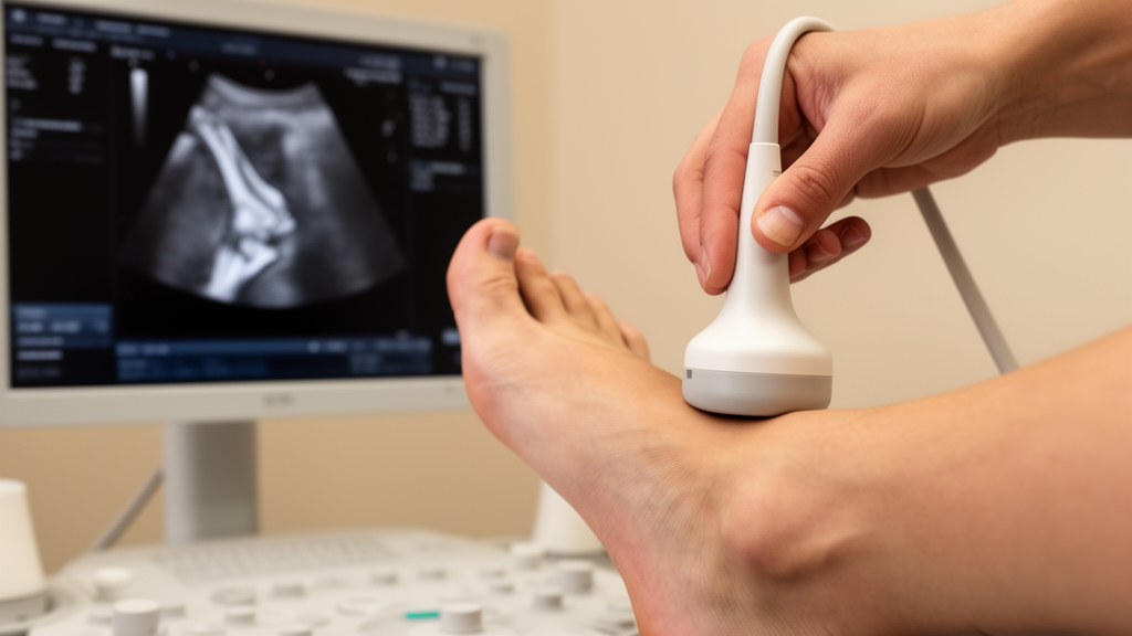
Ultrasonography can pinpoint where the peroneus brevis tendon attaches to the base of the fifth metatarsal with an average error of less than half a millimeter, according to a preliminary cadaveric validation study published in Discover Imaging on 4 August 2026. The study, led by Yuta Maeda and colleagues, tested whether a standard portable ultrasound machine could quantify the locations of two structures that anchor into the fifth metatarsal base: the peroneus brevis tendon and the lateral band of the plantar fascia. Both structures are thought to play a role in the biomechanics of fifth metatarsal stress fractures, known as Jones fractures, and the researchers wanted to establish whether ultrasound could measure their attachment points accurately enough to be used in living people.
The rationale for the work lies in a long-standing gap between anatomy and clinical practice. The peroneus brevis tendon and the lateral band of the plantar fascia both insert into the base of the fifth metatarsal, generating bending and torsional stress at the proximal portion of the bone, where Jones fractures occur, with the fourth/fifth intermetatarsal joint acting as the pivot point. Theoretical work suggests that differences in tendon attachment location change the bending stresses the bone experiences, and previous cadaveric studies have reported that the insertions of the peroneus brevis and the lateral band of the plantar fascia vary widely among individuals. That variation has been proposed as a possible, previously unidentified risk factor for Jones fractures, which are relatively uncommon but notorious among basketball and soccer players for prolonged absences from play, high rates of delayed union and non-union, and refracture after return to sport.
Measuring these attachment points in living athletes has been technically difficult. Because the peroneus brevis tendon and the lateral band of the plantar fascia insert in close proximity at the base of the fifth metatarsal, accessory slips and partial contiguity between the two structures can complicate precise discrimination. Magnetic resonance imaging and computed tomography have been shown to depict the locations of the two insertions reliably, but a previous imaging study did not analyze their quantitative validity. Ultrasound, by contrast, offers non-invasive, high-resolution imaging of tendon attachments and has already achieved millimeter-level accuracy in quantifying the dimensions of the Achilles tendon insertion. What remained unknown was whether the same precision was achievable for tendon insertions on a metatarsal bone.
To answer that question, the team used six fresh-frozen cadaveric feet, drawn from four Japanese cadavers with a mean age of 85.8 years, all male. Eight limbs were initially prepared, but two feet from one cadaver were excluded because of skin necrosis on the lateral aspect of the feet, leaving six feet comprising paired right and left feet from three cadavers. None of the specimens had a history of foot or ankle surgery or apparent deformity, and the absence of osteoarthritis was confirmed with fluoroscopy.
The measurement protocol was deliberately rigorous. Using a portable ultrasound machine with a high-frequency linear probe, the sonographer first defined the longitudinal axis of the fifth metatarsal by imaging the dorsolateral aspect of the bone in short axis, marking the midpoint of the flat portion of the cortical surface and a second point at the most distal portion of the metaphysis. Metal pins, 1.2 millimeters in diameter, were inserted vertically into the marked landmarks so that the same axis could be reconstructed after dissection. Next, the distal end of the fourth/fifth intermetatarsal joint was identified by scanning from proximal to distal until the intermetatarsal ligament disappeared and the dorsal articular contact ceased, the point at which the ultrasound beam penetrated deeper. Finally, the distal ends of the peroneus brevis and plantar fascia insertions were located by pivoting the probe around the relevant bony ridges until the fibrillar pattern of each structure was most clearly visualized, a maneuver designed to minimize anisotropy, the artifact that arises when ultrasound waves strike tendon fibers at an oblique angle.
Distances were then measured with a digital caliper in 0.1-millimeter increments along the fifth metatarsal axis, from the distal end of the fourth/fifth intermetatarsal joint to the distal ends of each insertion, with positive values assigned when a tendon insertion lay proximal to the joint. The sonographic examination was performed by an orthopedic fellow with two years of experience in musculoskeletal ultrasonography, including 100 cases of ultrasonographic screening for Jones fractures, and each measurement was performed twice with the mean used for analysis. After the imaging phase, a board-certified foot and ankle surgeon with 25 years of experience, blinded to the sonographic results, dissected each foot and repeated the identical measurements under direct visualization, defining the joint margin and the distal ends of the tendon footprints independently of the metal pins. The discrepancies between the two methods served as the primary outcome.
The results split cleanly between the two structures. The peroneus brevis tendon was correctly identified in all six feet, and the measurement error for its distance from the intermetatarsal joint was 0.3 plus or minus 1.5 millimeters, with a maximum error of 2.1 millimeters. The lateral band of the plantar fascia proved more difficult: it was misidentified in one foot, in which the most plantar fiber of the peroneus brevis was mistaken for the fascia, yielding a correct identification rate of five out of six feet. The overall measurement error for the fascia was 2.5 plus or minus 3.4 millimeters, but this fell to 1.2 plus or minus 1.6 millimeters when the misidentified foot was excluded. Under direct visualization, the peroneus brevis inserted more dorsally and the fascia more plantarly in all specimens, with their footprints partially contiguous at the proximal portions but clearly distinct at their distal ends.
The reference landmark itself proved highly reliable. The distal end of the fourth/fifth intermetatarsal joint, identified sonographically using the intermetatarsal ligament and dorsal articular contact, showed a positional deviation of only minus 0.1 plus or minus 0.9 millimeters relative to direct visualization, with a maximum deviation of 1.6 millimeters. The peroneus brevis insertion showed similarly minimal deviation, at minus 0.3 plus or minus 0.6 millimeters. The lateral band of the plantar fascia again showed greater variability, with a mean deviation of minus 1.0 millimeter that improved to 0.5 plus or minus 1.7 millimeters after exclusion of the misidentified case. The authors attribute the fascia’s difficulties not to the intrinsic resolution of ultrasound but to the anatomical complexity and partial contiguity of the two footprints, which insert in close proximity at the metatarsal base.
The accuracy achieved for the peroneus brevis carries particular weight because it was smaller than the magnitude of interindividual anatomical variability reported in previous cadaveric investigations of the same insertion. In other words, the measurement method is now precise enough to distinguish genuine anatomical differences between people from the noise of the measurement itself. This matters for Jones fracture research because small differences in insertion location may alter the effective moment arm and the stress distribution along the metadiaphyseal region of the bone. Previous clinical studies have focused on global alignment factors such as forefoot adduction, hindfoot varus, and restricted hip internal rotation as risk factors, and the new method provides the foundation to investigate a previously underexplored intrinsic factor: individual variation in tendon insertion anatomy, assessed non-invasively and at scale in athlete populations.
The authors are careful to frame the study as preliminary. It used elderly Japanese male cadaveric specimens, whose tissue quality, soft-tissue thickness, and degenerative changes may differ from those of young living athletes, the intended target population. Only six feet from three cadavers were analyzed, and because paired right and left feet came from the same donors, the observations may not be fully independent, a clustering effect that could not be statistically accounted for in the descriptive analysis. Sonographic measurements were performed by a single examiner, so intraobserver and interobserver reliability could not be evaluated, and the influence of examiner experience on accuracy remains unclear. Accessory bands of the peroneus brevis tendon, which may contribute to local stress around the fifth metatarsal base, were not evaluated because they could not be reliably identified by ultrasound, and footprint width or area was not assessed. The study also did not test whether variation in insertion anatomy is actually associated with Jones fractures. Even so, the authors conclude that ultrasound offers advantages over computed tomography and magnetic resonance imaging, including cost-effectiveness and feasibility for field-based studies, and that with refined scanning protocols, specific operator training, and validation in larger samples, the technique could become a practical tool for identifying individual anatomical risk factors for fifth metatarsal stress fractures in vivo.
Subject of Research: Validation of ultrasonography for measuring peroneus brevis and plantar fascia insertion locations at the fifth metatarsal base in cadaveric feet
Article Title: Accuracy of ultrasonography for quantifying tendon insertion locations at the fifth metatarsal base in a preliminary cadaveric validation study
Article References: Maeda, Y., Yamaguchi, S., Edama, M., Saita, Y., Suzuki, T., & Yamada, Y. (2026). Accuracy of ultrasonography for quantifying tendon insertion locations at the fifth metatarsal base in a preliminary cadaveric validation study. Discover Imaging, 3(1), Article 6. https://doi.org/10.1007/s44352-026-00029-2
Image Credits: AI Generated
DOI: 10.1007/s44352-026-00029-2
Keywords: ultrasonography, peroneus brevis, plantar fascia, fifth metatarsal, Jones fracture, cadaveric study, tendon insertion, fourth/fifth intermetatarsal joint, stress fracture, musculoskeletal imaging, biomechanics, validation study
News Source: Ophelia Keating. (October 28, 2026). Ultrasound Maps Tendon Anchor Points on the Fifth Metatarsal With Millimeter Accuracy. Scienmag.