In a groundbreaking advancement poised to reshape the landscape of liver cancer treatment, researchers at UCLA have embarked on an ambitious project to merge the capabilities of artificial intelligence with cutting-edge imaging technologies. Dr. Jason Chiang and Dr. Kyung Sung, eminent figures from the Department of Radiological Sciences at the David Geffen School of Medicine and UCLA Health Jonsson Comprehensive Cancer Center, recently secured a pivotal $3.2 million grant from the National Cancer Institute. This funding will fuel a five-year endeavor to craft an AI-enhanced imaging platform that aims to revolutionize the planning of yttrium-90 (Y90) radioembolization therapy for liver cancer patients.
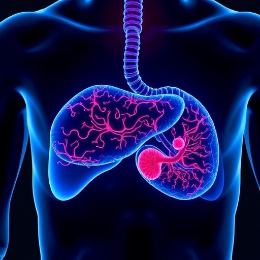
Y90 radioembolization represents a sophisticated, minimally invasive intervention uniquely tailored for targeted liver cancer treatment. The procedure involves the direct infusion of microspheres—microscopic radioactive beads—into hepatic tumors via the liver’s intricate vascular network. These microspheres lodge themselves within the tumor’s microvasculature, emitting localized radiation that eradicates malignant cells while preserving adjacent healthy liver tissue. The precision of this treatment stems not only from its direct delivery but also from meticulous calibration of the radioactive dose and bead dispersion. The complexity lies in achieving an optimal concentration of microspheres reaching the tumor; insufficient quantities fail to deliver therapeutic radiation levels, whereas excessive accumulation risks vascular occlusion and radiation spillover into non-target tissues, compromising efficacy and patient safety.
A critical bottleneck in current Y90 radioembolization planning revolves around the limitations of traditional imaging modalities in accurately predicting microsphere distribution. Despite advances in diagnostic imaging, the dynamic and heterogeneous nature of tumor blood flow poses a formidable challenge. Existing standard-of-care imaging fails to fully resolve the intricate and often erratic perfusion patterns within hepatic tumors, thereby impeding precise dosimetric calculations and bead deployment strategies. Recognizing this gap, Chiang—who also contributes his expertise to the UCLA Broad Stem Cell Research Center—and Sung, an AI and magnetic resonance imaging (MRI) savant, have synergistically combined their skill sets to pursue a novel strategy. Their approach integrates artificial intelligence with dynamic contrast-enhanced MRI (DCE-MRI), capturing the temporal and spatial variations in tumor vascularity with unprecedented fidelity.
By leveraging AI algorithms trained to interpret complex vascular patterns revealed by DCE-MRI, their platform aspires to generate predictive models that simulate Y90 microsphere behavior within liver tumors. This process involves dissecting the nuances of arterial blood flow, catheter placement during radioembolization, and tumor-specific vascular structures to forecast microsphere distribution precisely. The team’s rigorous methodology commences with the employment of specialized hepatic vascular phantoms—engineered physical models that mimic the liver’s vasculature and tumor microenvironment. These phantoms serve as controlled experimental arenas where investigators systematically analyze how alterations in arterial flow dynamics and catheter positioning influence microsphere dispersal.
Subsequent phases of development will extend validation studies to large animal models harboring liver tumors, utilizing clinically relevant MRI scanners and imaging protocols that mirror human clinical conditions. This translational approach ensures that findings derived from phantom studies gain biological relevance and are adaptable to real-world clinical scenarios. The integration of AI-powered image analysis with these experimental models aims to culminate in a robust computational toolkit capable of delivering personalized, high-precision planning for radioembolization therapy.
This pioneering blend of AI and advanced imaging embodies a significant stride toward personalized medicine in oncology. By deciphering and predicting the complex vascularity and microsphere dynamics within hepatic tumors, the platform promises to optimize therapeutic radiation delivery, minimizing deleterious off-target effects and enhancing overall patient outcomes. Such advancements could translate into higher tumor control rates, lower complication risks, and more refined treatment regimens tailored to individual tumor profiles.
Dr. Chiang emphasizes the transformative potential of this research, stating that the convergence of dynamic MRI techniques and artificial intelligence offers a new frontier in understanding microsphere distribution heterogeneity. This enhanced predictive power could empower clinicians to move beyond heuristic dosing paradigms toward data-driven, patient-specific treatment frameworks. Meanwhile, Dr. Sung highlights the AI system’s ability to process vast datasets encompassing temporal contrast dynamics and vascular geometries, thus unveiling patterns imperceptible to human interpretation.
The implications of this research extend beyond liver cancer treatment alone. The methodology developed—combining AI with dynamic contrast imaging—could inspire similar approaches in managing other solid tumors characterized by complex blood flow patterns. Moreover, success in this domain could catalyze new standards for integrating AI into interventional radiology, paving the way for real-time therapeutic planning and adaptive treatments.
This project stands as a testament to the power of interdisciplinary collaboration, melding expertise in clinical oncology, radiology, biomedical engineering, and artificial intelligence. The National Cancer Institute’s substantial investment underscores the high priority accorded to innovating cancer treatment modalities that improve efficacy while reducing adverse effects.
As the research unfolds over the coming years, the scientific and medical communities eagerly anticipate data that could substantiate the clinical benefits of AI-enhanced imaging platforms. Such breakthroughs may ultimately shift the paradigm of liver cancer treatment from empirical practices to precision-guided interventions, thus elevating standards of care and patient survival rates.
In conclusion, the pioneering work by Dr. Chiang, Dr. Sung, and their team illuminates the profound potential of integrating artificial intelligence with advanced imaging techniques. Their initiative promises to overcome longstanding obstacles in Y90 radioembolization planning, offering renewed hope for liver cancer patients worldwide. By harnessing cutting-edge technology and sophisticated computational modeling, this research heralds a new era of personalized, effective, and safer cancer therapies.
Subject of Research: Development of AI-enhanced imaging to optimize yttrium-90 radioembolization treatment planning for liver cancer.
Article Title: UCLA Researchers Secure $3.2M Grant to Revolutionize Liver Cancer Therapy with AI-Enhanced Imaging.
News Publication Date: Not specified.
Web References:
Dr. Jason Chiang Profile
Dr. Kyung Sung Profile
UCLA Health Jonsson Comprehensive Cancer Center
UCLA Broad Stem Cell Research Center
Keywords: Liver cancer, Yttrium-90 radioembolization, Artificial intelligence, Dynamic contrast-enhanced MRI, Tumor vascularity, Precision medicine, Computational modeling, Interventional radiology, Personalized cancer therapy, Hepatic vascular phantoms.
Tags: advanced cancer treatment technologiesAI in medical imaging and therapy planningAI-driven liver cancer treatmentAI-enhanced imaging platformminimally invasive cancer interventionsNational Cancer Institute grantpersonalized radioembolization therapyprecision oncology for liver tumorsradiological sciences in cancer caretargeted hepatic tumor therapyUCLA liver cancer researchyttrium-90 microsphere therapy
