Credit: Darling Lab / Brown University
PROVIDENCE, R.I. [Brown University] — A new technique developed by Brown University researchers reveals the forces involved at the cellular level during biological tissue formation and growth processes. The technique could be useful in better understanding how these processes work, and in studying how they may respond to environmental toxins or drug therapies.
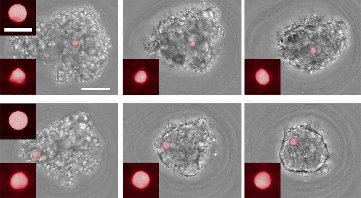
As described in the journal Biomaterials, the technique makes use of cell-sized spheres made from a highly compliant polymer material, which can be placed in laboratory cultures of tissue-forming cells. As the tissue-formation process unfolds, microscope imaging of the spheres, which are stained with fluorescent dye, reveals the extent to which they are deformed by the pressure of surrounding cells. A computational algorithm then uses that deformation to calculate the forces at work in that cellular microenvironment.
“We know that mechanical forces are important stimuli in tissue formation and development, but actually measuring those forces is pretty difficult,” said Eric Darling, an associate professor of medical science, engineering and orthopedics at Brown. “These spheres that we’ve developed give us an extremely sensitive technique for measuring those forces over time in the same sample. And we can do this with multiple samples at a time on a 96-well plate, so it’s a high-throughput method as well.”
The research was a collaboration between Darling’s lab and the lab of Haneesh Kesari, an assistant professor of engineering at Brown and an expert in solid mechanics. Darling and graduate student Robert Gutierrez developed the spheres and performed cell culture experiments with them, while Kesari and graduate student Wenqiang Fang developed the computational algorithm to calculate the forces.
The spheres are made from a polymer called polyacrylamide. The spheres have no apparent effect on the behavior of the newly forming tissues, Darling said, and the polyacrylamide material has mechanical properties that are highly consistent and tunable, which made it possible to make spheres soft enough to deform measurably when exposed to cellular forces.
“The key to this is having a highly controlled material, with a very precise shape as well as finely tuned and uniform mechanical stiffness,” Kesari said. “If we know the properties of the spheres, then we can take pictures of how their shapes change and back out the forces necessary to make those changes.”
As a proof of concept, the researchers performed a series of experiments to measure forces involved in mesenchymal condensation — a process in which stem cells cluster together and eventually differentiate into tissue-specific cell types. The process is central to the formation of teeth, bones, cartilage and other tissue.
In one experiment, the team included the force-sensing spheres in cultures of cells were coming together to form multicellular balls. Microscope images of the cultures were taken every hour for 14 hours, enabling the team to track changes in the forces involved in each culture over time. The experiments showed that the forces involved in mesenchymal condensation were highly variable for the first 5 or so hours of the process, before settling down into a much steadier force profile. This was the first time such force dynamics had ever been measured, the researchers say.
To help verify that the spheres were truly sensitive to cellular forces, the team repeated the experiment using cultures treated with a cytoskeletal inhibitor, a drug that weakens the tiny contractile motors inside a cell. As expected, the spheres detected markedly weaker forces in the cultures treated with the drug.
In another set of experiments, the researchers added the sensor spheres to preformed cellular masses to observe how the spheres were taken up into the mass. Some of the spheres had been treated with a collagen coating, which enables cells to bind with the sensors, while others were uncoated.
“We were able to see differences in the force profiles between the coated and uncoated spheres,” Darling said. “Overall there was a large compressive force, but with the coated cells we could see the cells interacting with the spheres directly, pulling on them and exerting a tensile force as well.”
Darling says he’s hopeful the technique could reveal fundamental details about how tissue-forming processes work. In the future, it may also be used screen drugs aimed at modulating these processes, or to test the effects of environmental toxins. It could also be useful in tissue engineering.
“If we want to grow cartilage, it might be helpful to know that the types of forces that these cells are exerting on each other because we might be able to apply an external force that matches or complements that force profile,” Darling said. “So in addition to fundamental discovery, I think there is some translational potential for this down the road.”
###
The work was funded by the National Institutes of Health (R01 AR063642), National Science Foundation (2018260690) and a Brown University Research Seed Award.
Media Contact
Kevin Stacey
[email protected]
Original Source
https:/
Related Journal Article
http://dx.
