Every living cell carries a hidden optical signature. The cytoplasm, the nucleus, and even the surrounding fluid each bend light by a slightly different amount, a property physicists call refractive index. Cancer cells, whose internal architecture differs from that of healthy cells, bend light differently too. A new computational design study, published in the Journal of Materials Science by Shivesh Kumar, Haraprasad Mondal, and Mrinal Sen, describes a photonic crystal nanocavity biosensor engineered to read precisely that signature, potentially offering a label-free route to discriminating cancer cells on a chip smaller than 83 square micrometers.
The device belongs to a family of structures known as photonic crystals, periodic arrangements of dielectric materials that control the flow of light in ways natural materials cannot. The concept, famously described by Joannopoulos, Villeneuve, and Fan in Nature in 1997, relies on a photonic bandgap, a range of wavelengths that the periodic structure simply refuses to transmit. When a defect is introduced into the otherwise perfect lattice, light of a specific wavelength becomes trapped at that defect, forming a nanocavity that acts like an extremely selective optical resonator.
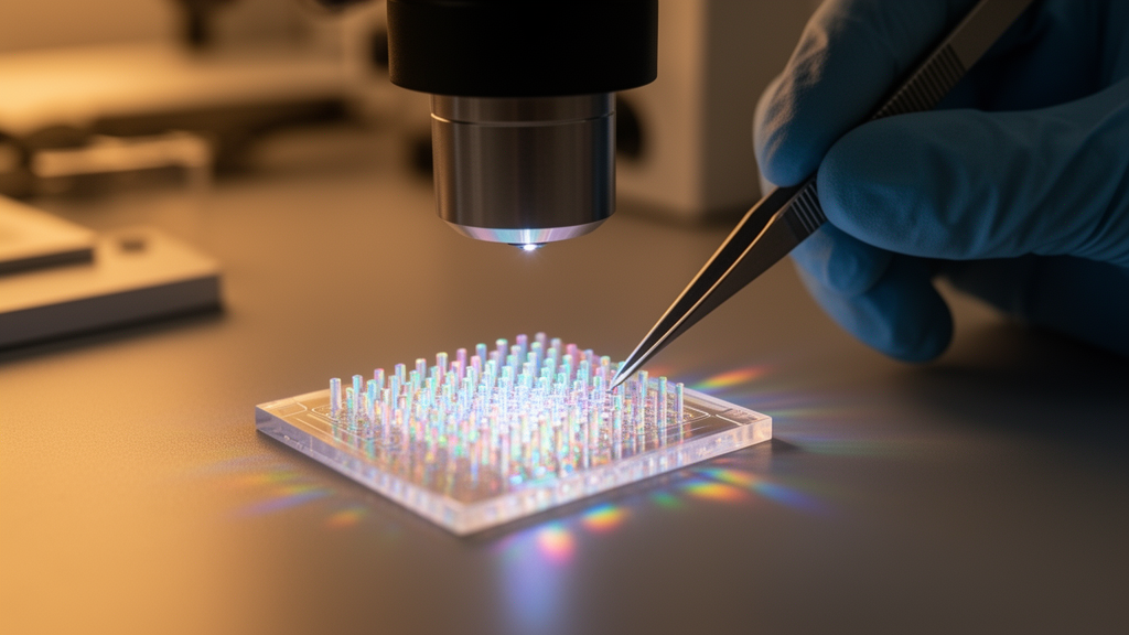
The proposed biosensor is based on a two-dimensional rod-in-air photonic crystal arranged in a triangular lattice configuration. Silicon rods stand in air, and the periodic arrangement creates a wide photonic bandgap with strong optical confinement within the sensing region. When a biological sample with a particular refractive index is placed in the cavity, the trapped resonance shifts to a new wavelength. By measuring that shift, the sensor can determine the refractive index of the sample with extraordinary precision, and because different cell types have different refractive indices, the same principle can be used to discriminate cancer cells from healthy ones.
The design process was systematic and rigorous. The photonic band structure was evaluated using the plane-wave expansion method, a computational technique that determines which wavelengths the periodic structure can and cannot transmit. Sensor performance was then analyzed through finite-difference time-domain simulations, which model how light actually behaves in the device over time. The researchers varied the rod radius within the sensing region at a constant lattice parameter, followed by an analysis of lattice constant variations while maintaining the optimized rod radius. Both two-dimensional and three-dimensional configurations were explored to validate the robustness of the design.
The performance figures are remarkable. For the fixed lattice constant configuration, the sensor achieves a sensitivity of 478.57 nanometers per refractive index unit, a quality factor of 1.6230 × 10^7, a detection limit of 1.91922 × 10^-8 refractive index units, and a figure of merit of 5.21043 × 10^6 per refractive index unit. In comparison, the variable lattice constant design provides a higher sensitivity of 965 nanometers per refractive index unit, along with a quality factor of 9.5621 × 10^5, a detection limit of 2.13245 × 10^-7 refractive index units, and a figure of merit of 4.68944 × 10^5 per refractive index unit.
These numbers deserve unpacking. Sensitivity describes how much the resonance wavelength shifts per unit change in refractive index, so a higher sensitivity means the device responds more dramatically to the presence of a biological sample. The quality factor, often abbreviated as Q, describes how sharply the resonance is defined, and a higher Q means the device can distinguish between very similar refractive indices. The detection limit describes the smallest refractive index change the device can reliably detect, and the figure of merit combines sensitivity and quality factor into a single performance score. The fixed lattice constant design achieves an extraordinarily high Q, while the variable lattice constant design achieves nearly double the sensitivity.
The practical challenges associated with photonic crystal fabrication were comprehensively analyzed. Photonic crystals require extremely precise periodic structures, and even small deviations from the ideal geometry can degrade performance. The researchers systematically investigated the impact of structural imperfections on sensor performance, providing a realistic assessment of how the device would behave in a laboratory setting. This kind of analysis is essential for translating computational designs into practical devices, and it distinguishes this work from purely theoretical studies that ignore fabrication realities.
The compact footprint of 82.84 square micrometers is a key advantage. On-chip photonic systems require devices that can be integrated densely, and the small size of this biosensor makes it a promising candidate for integration into advanced on-chip photonic systems. The device could potentially be combined with microfluidic channels that deliver biological samples directly to the sensing region, creating a complete diagnostic platform on a single chip.
The principle of refractive-index-based cancer cell discrimination is grounded in established biology. Cell nuclei have lower refractive index and mass density than cytoplasm, as demonstrated by Schürmann and colleagues in 2016, and cancer cells differ from healthy cells in their internal composition and organization. By measuring the refractive index of a cell with sufficient precision, the device can potentially identify whether that cell is cancerous. The approach is label-free, meaning no chemical tags or fluorescent markers are required, which simplifies the diagnostic process and avoids potential interference from labeling chemicals.
This work builds on a growing body of research into photonic crystal biosensors for biomedical applications. Previous studies have explored photonic crystal platforms for glucose detection, blood component analysis, urine biomarker detection, and cancer cell identification. The current design distinguishes itself through its combination of high quality factor, high sensitivity, low detection limit, and compact footprint, along with its realistic assessment of fabrication imperfections. The researchers, based at the Indian Institute of Technology (Indian School of Mines) Dhanbad and Dibrugarh University, acknowledge the Department of Electronics Engineering at IIT (ISM) Dhanbad for providing the research facilities and computational resources necessary to carry out this work.
Subject of Research: Computation & theory
Article Title: Design of a high-Q photonic crystal nanocavity biosensor for refractive-index-based cancer cell discrimination
Article References: Kumar, S., Mondal, H., & Sen, M. (2026). Design of a high-Q photonic crystal nanocavity biosensor for refractive-index-based cancer cell discrimination. Journal of Materials Science, 61(42), 32962-32984. https://doi.org/10.1007/s10853-026-13745-1
Image Credits: AI Generated
DOI: 10.1007/s10853-026-13745-1
Keywords: photonics, biosensors, cancer detection, nanocavities, silicon photonics, refractive index sensing, Design, high-Q, photonic, crystal, nanocavity, biosensor
News Source: Nathaniel Bowman. (October 18, 2026). Tiny Photonic Crystal Chip Could Tell Cancer Cells Apart by How They Bend Light. Scienmag.