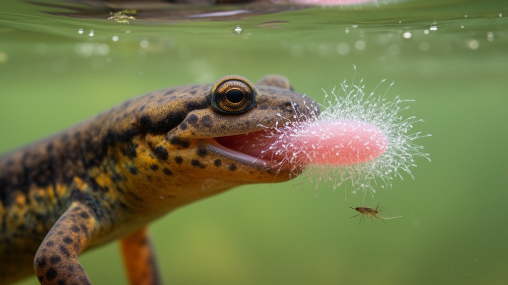
In the temporary ponds surrounding the German city of Jena, a population of smooth newts (Lissotriton vulgaris) has revealed a biological surprise that had gone unnoticed for as long as scientists have studied amphibian parasites. When researchers examined adult newts collected during their aquatic phase, they found the animals’ mouths densely colonized by sessile peritrich ciliates of the genus Apiosoma — single-celled protists that anchor themselves to surfaces with a stalk and sweep the water with a crown of cilia. The discovery, documented through histology and scanning electron microscopy, marks the first report of sessile ciliates infesting the oropharyngeal cavity of newts, and it raises intriguing questions about how such an invisible burden might undermine one of the most refined feeding mechanisms in the amphibian world.
The newts were collected in June 2017 from temporary ponds on the periphery of Jena, with permission from the city’s nature conservation authority. Two females and three males, all in their aquatic stage, were brought to the laboratory of the Zoological Institute of the University of Jena. The original aim of the study had nothing to do with parasites at all: the researchers wanted to understand how the feeding system of the species works. Only when the specimens were prepared for detailed anatomical analysis did the ciliates come to light, clinging in remarkable numbers to the lining of the animals’ mouths and throats.
To document the finding, the team employed two complementary techniques. For histological investigation, one male and one female newt were preserved in Bouin’s solution for two months, with the solution changed weekly. After decalcification, the samples were dehydrated in a graded ethanol-isopropanol series, embedded in paraffin, and cut into seven-micrometer semi-serial sections using rotatory microtomes. The sections were stained with periodic acid Schiff and a combination of hematoxylin-eosin, then examined under a light microscope. For scanning electron microscopy, three additional newts were processed: the floor of the mouth was removed and fixed in modified Karnovsky solution for eighteen hours, postfixed in osmium tetroxide, treated with dilute hydrochloric acid to wash mucus from the surface, dehydrated, critically point dried, gold-coated, and imaged in a scanning electron microscope.
Both methods revealed the same striking picture. The ciliate zooids were firmly attached to the oral epithelium and abundant throughout the oral cavity. The greatest concentration, roughly fifteen zooids per fifty square micrometers, occurred on the dorsal surface of the tongue. Under the scanning electron microscope, the ciliates appeared funnel-shaped, each with a narrow stalk anchoring it to the epithelial substrate and a broad peristomal disc fringed with numerous cilia. Horizontal annular striations, running perpendicular to the newt’s body axis, were clearly visible on the zooids. Histological sections showed granular structures within the ciliate bodies that stained positive for both alcian blue and periodic acid Schiff. Based on these morphological features, the researchers assigned the protists to the phylum Ciliophora, subclass Peritrichia, order Peritrichida, suborder Sessilina, genus Apiosoma.
Sessilid peritrich ciliates are no strangers to aquatic animals. They live as epibionts on a wide variety of aquatic invertebrates and also occur on vertebrates. Some species act as commensals, others as ectoparasites, and heavy infestations can even prove fatal to the host. Among amphibians, the best-documented peritrichs are the trichodinids, which inhabit the gills and skin of aquatic species and have occasionally been detected as endoparasites. Sessile peritrichs are well known as fish parasites, colonizing the body surface and gills. Historical accounts stretch back a century: as early as 1924, researchers reported various peritrich genera, including Vorticella, Opercularia, Rhabdostyla, and Epistylis, on the skin and gills of anuran larvae, some apparently restricted to the tadpole surface. In 1982, seven peritrich species were found on the skin of agile frog tadpoles, and one species, Apiosoma tintinnabulum, was recorded on the skin of the smooth newt itself.
What makes the new finding unusual is the location. Ciliates on skin and gills have been described many times, but a heavy infestation inside the oropharyngeal cavity of an amphibian is a rarely documented phenomenon. The researchers suggest such infestations may in fact be frequently overlooked, because the ciliates are invisible to the naked eye and the elaborate laboratory methods needed to detect them are rarely available in field studies. Without histology or electron microscopy, an infested newt would look perfectly normal even as its mouth harbored thousands of protists.
The ecological implications may be considerable, and here the study moves from observation to hypothesis. Smooth newts rely on water-dependent mechanisms for capturing prey and transporting it within the mouth during their aquatic phase. Like other vertebrates that feed by suction underwater, they lack prominent structures in the oropharynx, because anything protruding into the mouth can disrupt the rapid intraoral water flow on which suction feeding depends. Efficient suction feeding demands smooth, streamlined internal anatomy.
Smooth newts, in fact, display a remarkable form of phenotypic flexibility that makes this point vividly. During their aquatic season, their tongue surface is smooth, an adaptation thought to benefit underwater suction feeding. When they migrate to land, they grow a carpet of densely packed longitudinal lingual papillae, which presumably aids terrestrial feeding by helping grip prey. The seasonal transformation of the tongue is a textbook example of how morphology tracks ecological function. But the infested newts from Jena complicate this elegant picture: the ciliates covering their tongues were about the same size as the dorsal lingual papillae the animals would develop in their terrestrial stage. In effect, the protists had manufactured a papillae-like texture on a tongue that should have been hydrodynamically smooth.
The researchers hypothesize that this massive infestation functionally counteracts the newts’ sophisticated seasonal flexibility. A smooth tongue surface provides hydrodynamic benefits for suction feeding; cover it with numerous relatively large ciliates, and the water flows during suction may be disrupted. The densely packed zooids could cause drag within the waterflow in the bidirectional feeding situation characteristic of salamanders, and they may induce turbulent microflows within the lingual portion of the mouth. In practical terms, a newt whose tongue is carpeted with ciliates might suck water less efficiently, capture prey less successfully, and suffer reduced ecological performance — all without any visible sign of disease.
For fish, the consequences of external ciliate infestations are better understood: affected tissue on the skin and gills may produce excess mucus, and afflicted animals show loss of condition and characteristic behaviors such as flashing and piping. Researchers have also noted that the presence of numerous parasitic ciliates can signal deteriorated water quality, making these protists useful bioindicators. Whether the oropharyngeal Apiosoma of the Jena newts indicate something about the condition of their temporary ponds, or simply reflect an underappreciated and widespread phenomenon, remains open. What is clear is that a whole layer of amphibian–protist interaction may have been hiding in plain sight, inside the mouths of animals that scientists have studied for generations, visible only when the right microscopes are pointed at the right surfaces. As the authors conclude, an oropharyngeal cavity packed with ciliates is a phenomenon that deserves far more attention than it has ever received.
Subject of Research: Sessile peritrich ciliates of the genus Apiosoma colonizing the oropharyngeal cavity of smooth newts
Article Title: On the presence of the sessilid peritrich Apiosoma in the oro-pharynx of Lissotriton vulgaris ( Urodela , Vertebrata )
Article References: Natchev, N., & Heiss, E. (2026). On the presence of the sessilid peritrich Apiosoma in the oro-pharynx of Lissotriton vulgaris (Urodela, Vertebrata). Herpetozoa, 39, 23-26. https://doi.org/10.3897/herpetozoa.39.e173880
Image Credits: AI Generated
DOI: 10.3897/herpetozoa.39.e173880
Keywords: Apiosoma, smooth newt, Lissotriton vulgaris, peritrich ciliates, oropharynx, suction feeding, histology, scanning electron microscopy, amphibian parasites, phenotypic flexibility, tongue papillae, Germany
News Source: Drew Townsend. (October 17, 2026). Tiny Ciliates Carpet the Tongues of Smooth Newts, Threatening Their Suction Feeding. Scienmag.