
The intricate dance between myosin and actin is a cornerstone of cellular movement, a captivating process that invokes a deeper understanding of biomechanics at the molecular level. Recent advances in structural biology, particularly through the utilization of time-resolved cryo-electron microscopy, have unveiled exciting details about this relationship, specifically how myosin generates force and how actin catalyzes this movement. The fascinating interplay between these two proteins showcases the precision and elegance of cellular machinery.
In the realm of muscle contraction and cellular motility, myosin has long been recognized as a powerful motor protein. The mechanism by which it travels along actin filaments, primarily driven by ATP hydrolysis, is not only fundamental but also compelling. Studies have sequentially elucidated the structural transitions from unbound primed myosin to primed actomyosin and finally to the post-power stroke states. Each transition mirrors minute yet significant alterations in both the myosin and actin structures that facilitate this incredible force-generating capacity.
For instance, the preliminary weak binding of primed myosin to actin underlines a vital aspect of this interaction—the electrostatic complementarity between the positively charged residues of myosin’s loop 2 and the negatively charged residues in actin subdomain 1. This binding step is crucial as it positions the myosin lever arm in close proximity to the actin filament. These initial interactions trigger subsequent conformational changes within the myosin structure, setting the stage for force production.
As myosin engages actin, the interplay between the hydrophobic and ionic interactions leads to greater stabilization of the myosin’s light chain domain, referred to as L50. This stabilization is pivotal as it transitions myosin to the primed actomyosin state, characterized by a cocked position of the upper 50-kDa domain. The delicate balance of mechanical strain and molecular stabilization highlights the sophisticated choreography that occurs at the molecular level.
The transition from primed to the post-power stroke (postPS) state unveils the underlying principle of cleft closure in myosin. This mechanism is critically linked to the hydrolysis of ATP and results in a strong-binding interface necessary for enduring force production. As myosin experiences the lever swing, the transducer and relay helix undergo substantial reshaping, ultimately leading to the power stroke—a kinetic phenomenon observed in real-time through advanced imaging techniques.
Delving deeper into the mechanism of ATPase activation, one can observe how the N-terminal residues of actin play a crucial role. These residues become ordered upon binding with myosin, a structural change that significantly enhances actin’s ability to activate myosin’s ATPase activity. This rearrangement underscores the interdependence of myosin and actin as they engage in a dynamic partnership, critical for cellular movement.
Moreover, it is essential to highlight that while the actin structure remains relatively conserved through its various states, the motions of the N-terminal residues reveal a remarkable plasticity. This adaptability is central to the coordination required for efficient ATP hydrolysis and subsequent dissociation of inorganic phosphate (P_i). The rigorous timing of these events contributes to the overall efficiency of the power stroke.
The disassociation of P_i, a vital step catalyzed by the mechanical strain imparted by myosin actin binding, is a critical point of focus. The intricate dynamics showcase how myosin releases P_i in a carefully orchestrated fashion, illustrating the advantages of the energy economy present in cellular processes. This delay in phosphate release provides insights into how kinetic parameters govern the interaction dynamics, allowing for a seamless transition during muscle contraction.
Interestingly, the presence of an axial load introduces a layer of complexity in myosin’s functionality. Despite external resistance, myosin remains effectively coupled between cleft closure and lever swing, an observation that speaks to the motor protein’s ability to maintain its grip on actin filaments. Such resilience ensures that the force output remains consistent, underscoring how molecular mechanics operate with precision even under stress.
Fascinatingly, the serendipitous nature of these biochemical interactions illustrates a broader principle in molecular biology—the concept of cooperative binding. Both myosin and actin exemplify how slight changes in conformation can lead to significant functional outputs, a principle that resonates throughout numerous biological pathways.
The study of the myosin-actin interaction not only expands our comprehension of muscle physiology but also offers potential avenues in biotechnology and medicine. Understanding this mechanism can inspire new interventions in muscular disorders and provide groundwork for bioengineering applications where actin-myosin systems could be manipulated for desired outcomes.
Ultimately, the observations gleaned from this research provide a glimpse into the future of molecular biology and the vast potential for discovering novel therapeutic strategies and building synthetic biological systems. As our understanding of these molecular machines continues to evolve, we find ourselves at the threshold of groundbreaking discoveries that could transform how we approach biological function and disease.
By demystifying the mechanics underlying myosin and actin interactions, researchers are paving the way for innovations that promise to enhance our capabilities in various fields, from regenerative medicine to robotics. The journey of exploring these molecular interactions is just beginning, inviting further inquiry and exploration into the life-sustaining rhythms of cells.
In conclusion, myosin’s mechanism of movement, facilitated by actin catalysis, reflects the intricate balance of molecular forces that drive cellular dynamics. The continuous interplay between structure and function illustrates a profound aspect of life at the nanoscale, embodying the elegance and complexity that define biological systems. As we unravel these molecular mysteries, the impact of our discoveries holds the potential to redefine the interface between biology and technology, inspiring a new generation of scientists in their quest to understand and apply the wonders of life.
Subject of Research: Myosin and Actin Interactions in Muscle Contraction
Article Title: Swinging lever mechanism of myosin directly shown by time-resolved cryo-EM
Article References:
Klebl, D.P., McMillan, S.N., Risi, C. et al. Swinging lever mechanism of myosin directly shown by time-resolved cryo-EM. Nature (2025). https://doi.org/10.1038/s41586-025-08876-5
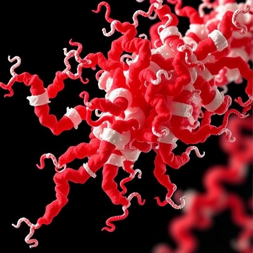
Image Credits: AI Generated
DOI: 10.1038/s41586-025-08876-5
Keywords: Myosin, actin, ATPase activation, muscle contraction, cryo-EM, molecular mechanics, cell motility, structural biology.
Tags: actin filament dynamicsATP hydrolysis in myosinbiomechanics at molecular levelcellular movement mechanismselectrostatic interactions in proteinsforce generation in cellsmotor protein functionmuscle contraction dynamicsmyosin actin interactionprotein structural transitionsstructural biology advancementstime-resolved cryo-electron microscopy

