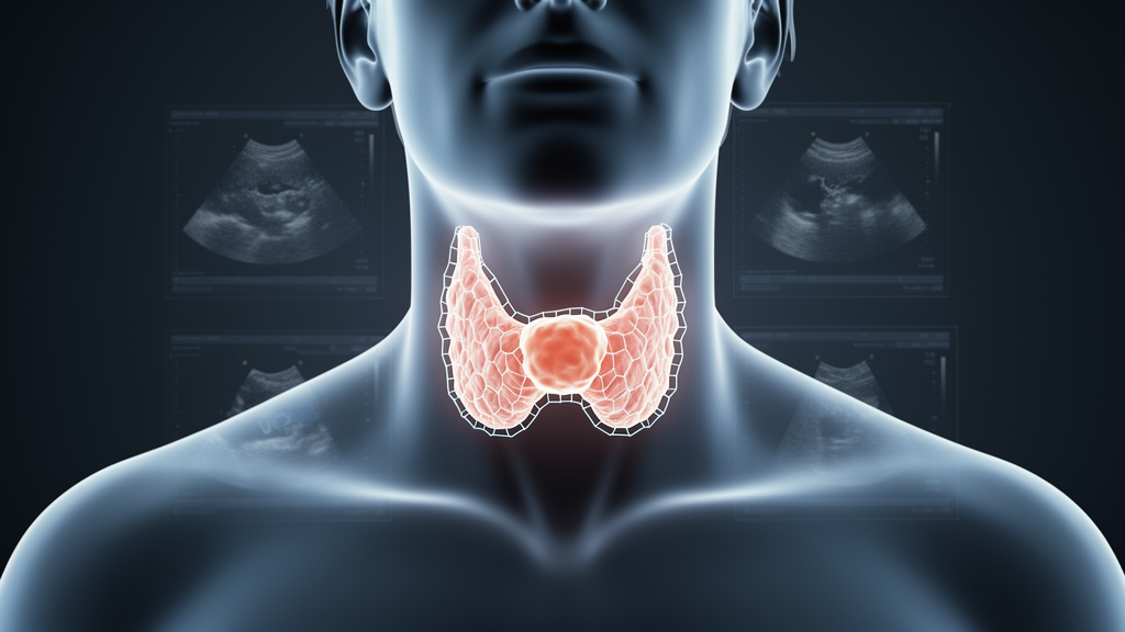
Artificial intelligence models that trace the outlines of thyroid nodules on ultrasound scans perform far less reliably when moved between medical centers, according to a comparative study published in the journal Cancer Imaging. Released online on 2 November 2026 as an open access article, the study benchmarked existing segmentation models under identical conditions and found that every model tested showed a consistent and substantial drop in accuracy across centers, with TNet achieving the best overall performance.
Segmentation models draw boundaries around thyroid nodules within an image, a step that determines what measurements a downstream diagnostic system can make. Because nodule size, shape and margin characteristics feed into risk assessment, an inaccurate boundary can distort clinical interpretation. The authors set out to compare the performance of existing thyroid nodule ultrasound segmentation models and to provide evidence for their clinical application, rather than to introduce a new architecture of their own.
To make the comparison fair, the team trained and evaluated all models under standardized conditions on two datasets. The first was the TN5000 public dataset, containing 5,000 images, which was split into training and internal validation sets. The second was an institutional dataset of 1,452 images, held back entirely and used as an independent external test set, so no model could tune itself to the external data.
TNet achieved the best overall performance, recording an external Dice score of 0.812. The Dice coefficient measures the overlap between the boundary a model draws and the boundary marked as correct, with 1 representing perfect agreement, so 0.812 indicates substantial but imperfect overlap on unseen external data. LGFFM showed higher recall, meaning it captured nodule regions while missing less of them, though it did not lead on overall overlap.
All models exhibited a consistent and substantial decline in performance over cross-center validation, so the loss of accuracy was not confined to one weak model. Non-thyroid-specific models performed worst, indicating that general-purpose architectures not tailored to thyroid ultrasound imagery fared poorly in this setting.
The authors concluded that existing models have limited cross-center generalizability, and that TNet performed best among the evaluated models in this experimental setting, a phrasing scoped to the specific datasets and conditions of the study. They stated that broader clinical adoption would require prospective multicenter validation before routine use could be justified. The work was published as a Research article under open access, and the journal notes that the early version is citable and carries a permanent DOI while remaining subject to further edits before the final Version of Record replaces it.
Subject of Research: Comparative evaluation of artificial intelligence models that segment thyroid nodules on ultrasound images
Article Title: Comparative evaluation of thyroid nodule ultrasound segmentation models
Article References: Li, C., Tang, Y., Wang, Y., Tan, M., Yao, L., Luo, Z., & Li, W. (2026). Comparative evaluation of thyroid nodule ultrasound segmentation models. Cancer Imaging. https://doi.org/10.1186/s40644-026-01149-y
Image Credits: AI Generated
DOI: 10.1186/s40644-026-01149-y
Keywords: thyroid nodule, ultrasound, image segmentation, deep learning, TNet, Dice score, cross-center validation, generalizability, TN5000 dataset, medical imaging, LGFFM, Cancer Imaging
News Source: Nathaniel Bowman. (November 7, 2026). Thyroid nodule AI segmentation models falter across medical centers. Scienmag.