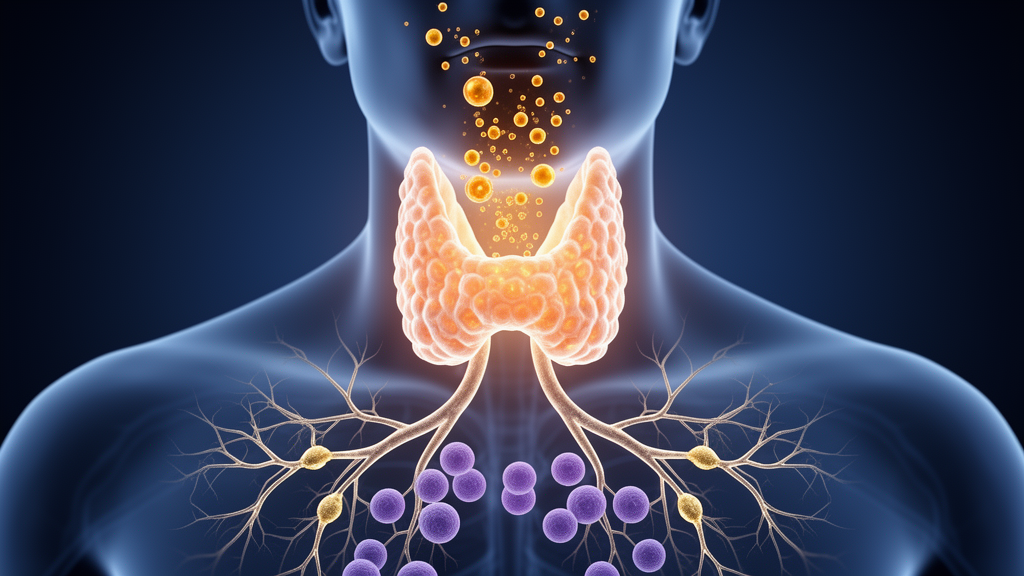
Scientists studying Parkinson’s disease have long suspected that the immune system and the thyroid gland are quietly entangled in the disorder, but a new retrospective study offers the clearest quantitative picture yet of where that entanglement holds and where it snaps. The research, published in npj Parkinson’s Disease, analyzed serum samples from 396 patients with Parkinson’s disease and 699 age- and sex-matched controls without thyroid disease, measuring a panel of thyroid hormones and inflammatory markers side by side. What emerged was a striking dissociation: the central command line linking the brain’s pituitary signals to the thyroid remained intact in Parkinson’s disease, while the peripheral machinery that converts thyroid hormones into their active forms appeared to be modulated by the immune system in a way that differs fundamentally from healthy physiology.
The study focused on four thyroid indices: thyroid-stimulating hormone, or TSH, which the pituitary releases to spur the thyroid gland; free thyroxine, or FT4, the main hormone the thyroid secretes; free triiodothyronine, or FT3, the more biologically active form produced largely by conversion of T4 in peripheral tissues; and the FT3/FT4 ratio, a practical readout of that peripheral conversion efficiency. On the immune side, the team measured C-reactive protein, an established marker of systemic inflammation, along with lymphocyte percentage, neutrophil percentage, and the neutrophil-to-lymphocyte ratio, a widely used composite index of inflammatory tone.
Group comparisons revealed selective rather than global thyroid disruption in Parkinson’s disease. Patients showed lower total T3, total T4, FT3, and the FT3/FT4 ratio compared with controls, while TSH and FT4 stayed within their reference ranges. That pattern matters because it points away from a primary failure of the thyroid gland itself or of the pituitary’s regulatory loop. If the central axis were broken, TSH would typically drift out of range alongside the hormones it governs. Instead, the preserved TSH and FT4 values alongside reduced FT3 and a lower conversion ratio suggest that something downstream, at the level of peripheral hormone conversion, is being reshaped in Parkinson’s disease.
The immune findings were equally distinctive. Patients with Parkinson’s disease had higher C-reactive protein levels, consistent with the chronic low-grade inflammation that has been documented in the disorder. Yet their differential blood counts told a more nuanced story: lymphocyte percentage was increased, neutrophil percentage was reduced, and the neutrophil-to-lymphocyte ratio was lower than in controls. This is not the classic inflammatory signature of acute infection, where neutrophils surge and lymphocytes recede. It is a remodeled immune balance, one that appears to coexist with the altered thyroid hormone profile rather than simply mirroring a systemic inflammatory flare.
The heart of the study lies in its correlation analyses, which asked how tightly thyroid indices and inflammatory markers track one another in each group. In controls, the FT3/FT4 ratio showed consistent associations with immune markers, indicating a stable physiological coupling between peripheral hormone conversion and immune status in healthy people. In Parkinson’s disease, those associations were attenuated and became dependent on immune status, meaning the strength and direction of the thyroid-immune relationship shifted depending on where a patient fell along the inflammatory spectrum. The researchers used partial correlation analyses, interaction models, and segmented regression to probe these relationships, allowing them to detect threshold effects and context-dependent coupling that simple linear correlations would miss.
This dissociation between central and peripheral axes provides a framework for understanding why thyroid findings in Parkinson’s disease have been so heterogeneous across prior studies. Some investigations have reported subclinical hypothyroidism, others normal thyroid function, and still others altered T3 levels without a clear mechanism. The new results suggest that these inconsistencies may reflect a context-dependent regulatory state: the central axis continues to operate, but the peripheral conversion of T4 to T3 is being modulated by immune signals in ways that vary from patient to patient. A patient with a particular inflammatory profile might show one pattern of thyroid indices, while another with a different immune balance might show an entirely different one, even though both have the same underlying neurodegenerative disease.
To test whether these associations generalize beyond the original clinic-based cohort, the team examined key relationships in an independent population-based sample from the United States drawn from the National Health and Nutrition Examination Survey. Replicating associations in a second, demographically distinct cohort strengthens the argument that the thyroid-immune decoupling observed in Parkinson’s disease is not an artifact of a single clinical population, referral pattern, or laboratory methodology. Population-based replication is particularly valuable in this field, where clinic-based studies can overrepresent patients with more advanced disease or comorbid conditions that independently affect both immune and thyroid measures.
The mechanistic implications reach into several active areas of Parkinson’s disease research. Chronic inflammation has been implicated in the progression of the disorder, with elevated C-reactive protein previously associated with worse motor outcomes and faster decline in some cohorts. Thyroid hormones, meanwhile, influence neuronal metabolism, mitochondrial function, and the clearance of misfolded proteins. If peripheral conversion of T4 to T3 is being modulated by immune status in Parkinson’s disease, then fluctuations in inflammatory tone could translate into fluctuations in the hormonal environment that neurons experience, adding an endocrine dimension to the well-established immune-neural crosstalk in the disorder. The study does not establish causation or direction, and the authors note that how immune status influences thyroid function, and at what level such effects emerge, remains an open question that the retrospective design cannot fully resolve.
What the study does establish is a measurable architecture. The preserved central axis activity alongside immune-linked modulation of peripheral hormone conversion is not a vague suggestion of endocrine involvement; it is a specific, testable pattern that future longitudinal studies can track over time. If the immune-dependent coupling of the FT3/FT4 ratio proves to change as Parkinson’s disease progresses, or if it responds to anti-inflammatory interventions, it could become a useful biomarker axis for monitoring the disorder’s systemic physiology. The finding that thyroid regulation in Parkinson’s disease is context-dependent rather than uniformly disrupted reframes the thyroid from a passive bystander into an active participant whose behavior is shaped by the immune milieu, offering a quantitative bridge between two biological systems that Parkinson’s disease research has largely treated separately.
Subject of Research: Thyroid-immune interactions and peripheral hormone conversion in Parkinson's disease
Article Title: Differential thyroid–immune coupling across central and peripheral axes in Parkinson’s disease
Article References: Feng, C., Zhang, Z., Xu, J., Cui, X., Zhou, X., & Xu, Y. (2026). Differential thyroid–immune coupling across central and peripheral axes in Parkinson’s disease. npj Parkinson's Disease. https://doi.org/10.1038/s41531-026-01570-x
Image Credits: AI Generated
DOI: 10.1038/s41531-026-01570-x
Keywords: Differential, thyroid, immune, coupling, across, central, peripheral, axes, Parkinson, disease, scientific research
News Source: Diana Fleming. (October 15, 2026). Thyroid Hormone Signals Shift in Parkinson’s Disease as Immune Coupling Breaks Down. Scienmag.