Skin cancer remains one of the most common and potentially deadly forms of malignancy worldwide, but its outcome depends dramatically on how early it is caught. A lesion identified in its initial stages is often curable with a simple excision, while the same lesion detected late can metastasize into a life-threatening disease. That is why dermatologists have long sought tools that can help them triage suspicious moles and spots quickly and reliably. Now, a team of researchers in India has unveiled a deep learning framework that fuses three different convolutional neural network architectures into a single, unified classifier, and the results are striking: accuracies approaching 99 percent on two of the most widely used dermoscopic image benchmarks in the field. The system, called SkinEnsemNet, was described in a study published in the journal Multimedia Tools and Applications, and it offers a compelling demonstration of how architectural diversity, rather than raw model size, can drive performance gains in medical image analysis.
The core idea behind SkinEnsemNet is deceptively simple. Instead of betting everything on one neural network, the researchers combined three pretrained convolutional neural network backbones that each see dermoscopic images through a different computational lens. The first, VGG16, is a classic deep architecture known for capturing localized spatial and hierarchical features through its stack of small convolutional filters. The second, DenseNet201, belongs to a family of densely connected networks in which each layer receives feature maps from all preceding layers, enabling extensive feature reuse and efficient hierarchical extraction. The third, EfficientNetB0, was designed to balance accuracy against computational cost through a systematic compound scaling of network depth, width, and resolution. By pooling the strengths of these three very different models, the framework aims to exploit complementary representations of the same skin lesion image, something no single architecture can fully achieve on its own.
The technical pipeline behind the system is worth unpacking, because it illustrates the careful engineering that underpins modern medical AI. The researchers began with transfer learning, meaning that each of the three backbones had already been trained on ImageNet, a massive general-purpose image database, before being adapted to dermoscopy. Transfer learning allows models to inherit rich visual features learned from millions of natural images, which is especially valuable when medical training data are limited. Before any image reached the networks, the pipeline applied normalization to standardize pixel values, and it tackled one of the most persistent problems in dermatological datasets: class imbalance. Some lesion categories are far more common than others, and naive models tend to ignore rare but clinically dangerous classes. To counter this, the team used random oversampling, duplicating examples from underrepresented classes until the training distribution was more balanced.
Once the three backbone networks had processed an image, their outputs were not simply averaged or voted on. Instead, SkinEnsemNet performs feature-level fusion. Global average pooling condenses each backbone’s high-dimensional feature maps into compact vectors, and these vectors are then concatenated into a single joint representation that carries spatial, hierarchical, and efficiency-oriented information simultaneously. On top of this fused representation sits a stack of dense classification layers, with dataset-specific heads allowing the same framework to be tailored to different benchmarks. Dropout regularization was applied throughout to prevent the network from memorizing training examples, a critical safeguard when the goal is generalization to unseen lesions. This design means the ensemble learns how to weigh and combine evidence from its three constituent models, rather than relying on a crude post-hoc vote.
The performance numbers reported in the study are impressive. On held-out image-level test partitions, SkinEnsemNet achieved an accuracy of 98.85 percent on the HAM10000 dataset, a large collection of multi-source dermatoscopic images of common pigmented skin lesions, and 97.83 percent on ISIC-2019, a broader benchmark spanning eight lesion categories. The corresponding weighted F1-scores, which balance precision and recall while accounting for class frequencies, were 98.86 percent and 97.83 percent respectively. Crucially, the ensemble outperformed each of the individual CNN backbones when those were evaluated under the same experimental protocol, confirming that the fusion strategy, and not merely the quality of any single model, was responsible for the gains. In a field where a fraction of a percentage point can mean the difference between a missed melanoma and a timely diagnosis, such consistent improvements carry real weight.
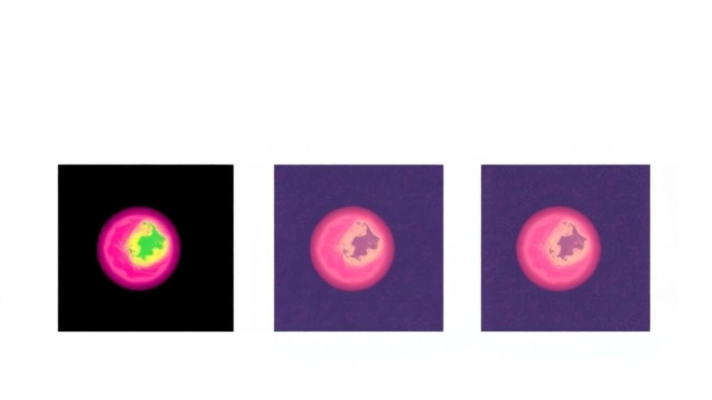
Accuracy alone, however, is not enough for a tool that might one day influence clinical decisions. A model that performs well but cannot explain why it reached a conclusion is unlikely to earn the trust of dermatologists. Addressing this, the researchers applied Grad-CAM, a gradient-based visualization technique that highlights the image regions most influential in a given prediction. The resulting heatmaps offer qualitative insight into whether the network is attending to the actual lesion boundaries, pigmentation patterns, and structural features that clinicians consider, or whether it is being distracted by irrelevant artifacts such as rulers, hair, or dark corners of the dermoscopic frame. While the visualizations are qualitative rather than a formal proof of correctness, they represent an important step toward interpretable AI in dermatology, where black-box predictions have historically been met with justified skepticism.
The choice of datasets deserves attention as well. HAM10000, assembled by Tschandl and colleagues, has become a de facto standard for skin lesion classification research, containing over ten thousand dermatoscopic images across seven diagnostic categories including melanoma, basal cell carcinoma, and benign nevi. ISIC-2019, drawn from the International Skin Imaging Collaboration, extends the challenge to eight classes and greater image diversity. Testing on both benchmarks, with dataset-specific classification layers, demonstrates that the framework’s architecture is not overfit to the quirks of a single data collection. That said, the authors themselves are careful to frame these results as strong within-dataset performance, a distinction that matters enormously in the translation of laboratory models to real-world clinics.
Indeed, the study’s authors are notably candid about the limits of what they have shown and the work that remains. They call for further evaluation using patient-level splitting, in which all images from a single patient are kept on the same side of the train-test divide, preventing the model from effectively memorizing individuals. They also recommend testing on external datasets collected at different institutions and with different imaging equipment, a rigorous test of generalization that within-dataset splits cannot provide. Beyond that, they point toward calibration analysis, which assesses whether the model’s confidence scores actually reflect the true probability of a diagnosis, as well as formal clinician assessment and deployment-oriented experiments. These are exactly the kinds of validation steps that regulatory bodies and medical professionals demand before an algorithm can move from a research paper to a hospital workstation.
The broader context makes this work part of a fast-moving wave. Deep learning has transformed medical image analysis over the past decade, and skin cancer detection has been one of its most visible battlegrounds, with convolutional networks repeatedly matching or exceeding expert dermatologists on curated benchmarks. Yet most published systems rely on a single architecture, leaving performance vulnerable to the blind spots of that particular design. Ensemble approaches that deliberately combine heterogeneous backbones, as SkinEnsemNet does, point to a strategy that trades some computational overhead for robustness and accuracy. With EfficientNetB0 already among the most computationally efficient architectures available, the authors suggest that such a framework could eventually run in settings where resources are constrained, from primary care clinics to smartphone-based screening tools in underserved regions.
For now, SkinEnsemNet stands as a rigorous proof of concept rather than a finished clinical product, but its trajectory is clear. The framework shows that thoughtfully orchestrated architectural diversity, disciplined preprocessing, and attention to interpretability can push dermoscopic classification to near-ceiling performance on standard benchmarks. The path from there to the clinic runs through external validation, calibration, and collaboration with the dermatologists whose judgment these tools are meant to augment, not replace. If those steps bear out the promise of the current results, the humble mole photographed through a dermatoscope may soon be scrutinized not just by one pair of expert eyes, but by a committee of three very different artificial minds, each catching what the others might miss, and together edging medicine closer to the goal of catching deadly skin cancers before they ever get the chance to spread.
Subject of Research: Ensemble deep learning for skin cancer classification from dermoscopic images
Article Title: SkinEnsemNet: an ensemble deep learning-based framework for the classification of skin cancer
Article References: Lodh, E., Chowdhury, T., Majumder, S., De, M., & Singha, S. (2026). SkinEnsemNet: an ensemble deep learning-based framework for the classification of skin cancer. Multimedia Tools and Applications, 85(9), Article 726. https://doi.org/10.1007/s11042-026-21892-5
Image Credits: AI Generated
DOI: 10.1007/s11042-026-21892-5
Keywords: skin cancer, deep learning, ensemble learning, transfer learning, VGG16, DenseNet201, EfficientNetB0, HAM10000, ISIC-2019, Grad-CAM, dermoscopy, convolutional neural networks
News Source: Nathaniel Bowman. (October 6, 2026). Three AI Models, One Verdict: Ensemble Network Pushes Skin Cancer Detection Toward 99% Accuracy. Scienmag.