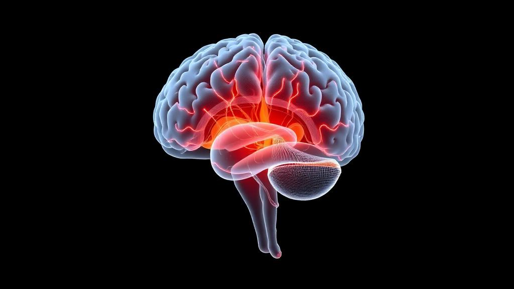
Restless legs syndrome has long resisted a single explanation. Brain iron deficits, dopamine signaling problems, glutamate and GABA imbalance, adenosine-related arousal mechanisms, and abnormal sensory processing have each been implicated, yet most research has examined them in isolation. A new hypothesis paper published in Frontiers in Human Neuroscience argues that these mechanisms may converge on a common network node: the thalamus, a deep brain structure that simultaneously handles sensory gating, motor-loop modulation, arousal regulation, and sleep-wake stability.
The article, authored by Beatrix Vághy of Semmelweis University, Sámuel Komoly of the University of Pécs, and Szabolcs Várbíró, does not claim that restless legs syndrome is a primary thalamic disorder. Instead, it proposes a thalamus-centered convergence model in which iron deficiency, dopaminergic instability, glutamatergic hyperresponsiveness, reduced adenosinergic inhibition, altered GABAergic modulation, and abnormal afferent input interact to make thalamo-cortical networks vulnerable to sensory and arousal instability. The authors frame the model as a way of organizing established frameworks within a unified, testable architecture rather than replacing them.
To motivate the model, the authors present three illustrative clinical phenotypes drawn from routine diagnostic care. The first is a 62-year-old man who developed symptoms at age 54 and was treated with pramipexole, which was escalated over several years without specialist follow-up to 1.08 mg per day. His evening discomfort crept earlier into the afternoon, spread from the calves toward the thighs, and became functionally disabling. Polysomnography showed a periodic limb movement index of 62 per hour and substantial sleep fragmentation, with normal breathing parameters. The clear temporal association with dose escalation supported dopamine-agonist augmentation, a treatment-related complication that contemporary guidance, including the 2025 American Academy of Sleep Medicine guideline, seeks to avoid by advising against long-term pramipexole, ropinirole, rotigotine, or levodopa.
The second phenotype is a 54-year-old man with sensory-predominant symptoms, an IRLS severity score of 26 out of 40, and relatively little overt motor activity. Pregabalin titrated from 150 to 300 mg per day over six months produced no meaningful improvement. His polysomnography showed a low periodic limb movement index of 14 per hour but pronounced sleep fragmentation and reduced N3 sleep, with normal respiratory measures. The authors note that repeated overnight testing was declined, so the sleep data reflect a single night. This case highlights the dissociation between motor burden and sensory symptoms, making it relevant to models centered on sensory processing and arousal regulation rather than periodic limb movements alone.
The third case involves a 32-year-old woman with moderately severe symptoms, an IRLS score of 27 out of 40, and peripheral iron deficiency attributed to heavy menstrual bleeding and suboptimal dietary intake. After three months of oral ferrous sulfate, her ferritin rose from 58 to 92 micrograms per liter and transferrin saturation from 17 to 24 percent, yet her symptoms barely improved, with the score falling only to 25 out of 40. The authors emphasize that these values represent partial correction rather than the iron repletion thresholds typically associated with symptom relief, and that intravenous ferric carboxymaltose, which the AASM guideline supports, was considered but deferred. The case illustrates that peripheral iron markers are imperfect surrogates for brain iron biology.
Despite their different biological contexts, the three cases converge on two broad outcomes: persistent sensorimotor discomfort and impaired sleep continuity. All three showed sleep efficiency around 66 to 68 percent and wake after sleep onset between 110 and 125 minutes, while their periodic limb movement indices ranged widely, from 14 to 62 per hour. The authors are careful about what this convergence demonstrates. The cases support only a modest inference, that distinct upstream contexts can lead to superficially similar clinical and sleep-physiological endpoints. They do not identify where in the nervous system convergence occurs, and because none of the patients underwent thalamus-specific MRI, spectroscopy, or neurophysiological measurement, no inference about individual thalamic dysfunction can be drawn from them.
The thalamic evidence comes instead from independent human studies. Proton magnetic resonance spectroscopy work reported higher thalamic glutamate-glutamine signal in restless legs syndrome and an association with wake time during the sleep period, without a corresponding relationship to periodic limb movement frequency. The authors treat this finding cautiously, noting that the underlying study examined right-sided thalamic spectroscopy in a relatively small sample of 28 patients and 20 controls, predates newer multimodal imaging literature, and has seen sparse independent replication. They also stress that the spectroscopy signal is a composite of glutamate and glutamine and does not directly measure synaptic glutamatergic firing. Functional connectivity studies have reported altered thalamic connectivity or network topology, and metabolic imaging has shown abnormal medial thalamic metabolism, while structural findings are less uniform, with some studies reporting gray-matter changes and others finding no consistent subcortical volumetric differences.
Within the proposed model, iron and dopamine act as upstream modulators rather than primary lesion sites. Iron availability affects dopaminergic signaling, glutamatergic-GABAergic balance, and metabolic efficiency, while dopamine modulates excitability, gain control, and network stability. The model also incorporates a hypoadenosinergic state as an indirect, network-level hypothesis, since much of that evidence derives from experimental and corticostriatal models rather than direct thalamic measurements. Sensory gating is described as a state-dependent, network-level process influenced by dopaminergic tone, iron-dependent neurotransmission, and circadian modulation, which could amplify otherwise low-salience limb-related input during evening and nighttime hours when external sensory competition is reduced.
The authors outline six falsifiable predictions to make the framework empirically accountable. These include testing whether multivariate thalamic connectivity measures explain variance in symptom severity and sleep fragmentation after adjustment for age, sex, medication exposure, and movement burden; whether thalamic glutamate-glutamine signal correlates more strongly with hyperarousal and sleep microstructure than with periodic limb movements; whether thalamic abnormalities are state-dependent and more pronounced in the evening; whether combining sensory and thalamic measures explains severity better than either alone; whether sleep spindle and cyclic alternating pattern measures mediate the link between severity and subjective sleep disturbance; and whether successful therapies acting through different pathways share partial normalization of thalamic biomarkers. The authors also flag lower-limb thalamic somatotopy as a speculative component, since no direct study has demonstrated selective dysfunction of lower-limb thalamic representations in the syndrome.
The paper is explicit about its limitations. The three phenotypes are illustrative rather than evidentiary, most thalamic findings are cross-sectional and associative and may be influenced by medication exposure, symptom state, and sleep deprivation, and the term thalamus encompasses heterogeneous nuclei with distinct functions that future work should resolve individually. The authors emphasize that the thalamus is central in their diagram because of its integrative position, not because every proposed link carries equal empirical support, and that cortico-striatal circuits, brainstem arousal systems, spinal mechanisms, and distributed sensory networks may contribute equally or more strongly in specific patient subgroups. Whether the model earns its place, they conclude, will depend on prospective testing: if thalamic network measures explain clinically meaningful variance beyond competing cortico-striatal, brainstem, or spinal models, the framework gains support, and if not, alternative convergence architectures should be favored.
Subject of Research: A thalamus-centered network model of restless legs syndrome integrating iron, dopamine, glutamate, GABA, adenosine and sensory mechanisms
Article Title: A thalamus-centered network model of restless legs syndrome: an integrative hypothesis illustrated by three clinical phenotypes
Article References: Vághy, B., Komoly, S., & Várbíró, S. (2026). A thalamus-centered network model of restless legs syndrome: an integrative hypothesis illustrated by three clinical phenotypes. Frontiers in Human Neuroscience, 20, Article 1977941. https://doi.org/10.3389/fnhum.2026.1977941
Image Credits: AI Generated
DOI: 10.3389/fnhum.2026.1977941
Keywords: restless legs syndrome, thalamus, thalamo-cortical networks, brain iron, dopamine agonist augmentation, glutamate, sensory gating, sleep fragmentation, periodic limb movements, magnetic resonance spectroscopy, hypoadenosinergic state, falsifiable predictions
News Source: Cassandra Pierce. (October 28, 2026). Thalamus Proposed as Convergence Hub in New Restless Legs Syndrome Model. Scienmag.