A multicentre survey of referral hospitals across 17 African countries has found that posterior urethral valves, the leading congenital obstruction of the lower urinary tract in boys, are still being diagnosed late and treated with limited surgical resources across much of the continent. The study, conducted by the group Modern Family for Urology in Africa (MUFA) and published in the African Journal of Urology, collected standardised questionnaires from urologists and paediatric surgeons and analysed responses from 31 centres that actively manage the condition. Its results paint a picture of fragmented care in which subspecialist expertise is scarce, prenatal detection is rare, and the endoscopic equipment needed for standard treatment is absent from a large share of institutions.
Posterior urethral valves are folds of tissue in the male urethra that obstruct the flow of urine from the bladder. Globally, the condition is estimated to affect between 1 in 5,000 and 1 in 25,000 live male births, although the authors note that its precise incidence in low- and middle-income countries remains unknown. The obstruction can cause obstructive uropathy and chronic kidney disease, and in sub-Saharan Africa the burden of disease is poorly understood because longitudinal data and specialised paediatric urological infrastructure are critically lacking. In high-resource settings, prenatal detection is standard; in the African centres surveyed, patients typically arrive during childhood with advanced urological complications, and referral services are dominated by the management of secondary sequelae such as persistent bladder dysfunction and renal failure.
The survey received 48 responses in total, of which 31 centres reported managing patients with posterior urethral valves and were included in the analysis. Nigeria was the most represented country with 10 centres, followed by Cameroon and Tanzania with three each and Ethiopia with two; the remaining 13 countries contributed one centre apiece. Organisationally, 23 centres, or 74.2 percent, had a dedicated paediatric surgery department, while 8 did not. When centres rated their own level of expertise in paediatric urology, 8 described it as low, 21 as moderate, and only 2 as high, meaning that 93.5 percent of centres rated their expertise as low or moderate. Hypospadias was the most commonly managed paediatric urological condition, reported by 24 of the 31 centres, with posterior urethral valves ranked among the three most frequently managed conditions at 19 centres.
The timing of diagnosis emerged as one of the starkest findings. The most frequent age at diagnosis was between 1 and 5 years, reported by 17 of the 31 centres, or 54.8 percent, while diagnosis before 1 year of age occurred at 10 centres and after 5 years at 4 centres. Prenatal diagnosis was described as rare by 18 centres, never available by 7, and frequent by only 6, meaning it was uncommon or absent in the overwhelming majority of institutions. This pattern is consistent with earlier regional reports cited by the authors: prenatal diagnosis was identified in only 11.3 percent of Sudanese cases and in none at a Nigerian hospital, and median ages at diagnosis have reached 2 years in Cameroon and as much as 5 years and 7 months elsewhere. Malnutrition, present in 36 percent of a Kenyan cohort, compounds these delays by contributing to high mortality and irreversible renal damage.
Diagnostic resources were unevenly distributed. Renal and urinary tract ultrasonography combined with CT scanning was the most widely available imaging modality, present at 25 of the 31 centres, or 80.6 percent. Voiding cystourethrography was used at 22 centres, or 71.0 percent, and cystoscopy at 19 centres, or 61.3 percent, when indicated. On the therapeutic side, 19 centres, or 61.3 percent, had on-site paediatric endoscopy facilities, while 12, or 38.7 percent, had none. Endoscopic valve ablation, the treatment generally regarded as the standard of care for posterior urethral valves, was performed at only 9 centres, or 29.0 percent, and endoscopic resection procedures were likewise available at 9 centres. Paediatric ureteroscopy was available at just 2 centres, and JJ stent placement for urinary drainage was reported at 6. The authors caution that centres without on-site facilities may still refer patients elsewhere for cystoscopy or definitive treatment, a pathway the survey did not separately capture.
Where specialised instrumentation is lacking, treatment often falls back on alternative techniques. The article notes that management in regional facilities frequently includes Mohan’s valvotomy, catheter balloon avulsion, and open resection alongside endoscopic approaches. In one 10-year Nigerian experience, Mohan’s valvotomy was the primary treatment for 47.6 percent of patients, compared with only 33.3 percent receiving endoscopic ablation. Clinical presentation in these settings is typically severe, with patients reporting poor urinary stream in 79.2 percent of cases, recurrent fever in 62.3 percent, and abdominal distension, and elevated serum creatinine observed in nearly 80 percent of patients at the time of diagnosis.
The consequences of late detection and restricted access to endourological care extend well beyond the operating theatre. A study in Ethiopia found that 52.9 percent of patients developed postoperative chronic kidney disease during three years of follow-up, with risk factors including postoperative hypertension and an elevated nadir serum creatinine. Bladder dysfunction also persists after treatment: at Kenyatta National Hospital, significant post-void residual urine was documented in 79 percent of patients following ablation. Loss to follow-up is another critical barrier, reported as high as 40 percent in some series, often driven by the mistaken belief among families that initial surgical relief of the obstruction constitutes a complete cure. Mortality rates ranging from 4 to 13 percent have been reported in some African cohorts, primarily related to urosepsis and advanced renal failure.
When asked about the main obstacles to optimal care, the centres ranked lack of endoscopic equipment first, cited by 14 of 31 centres, or 45.2 percent. The cost of care for families followed at 10 centres, or 32.3 percent, and limitations in technical facilities at 7 centres, or 22.6 percent. The authors observe that financial limitations are particularly significant in low-resource settings, where access to specialised investigations, surgery, and long-term follow-up often depends on out-of-pocket expenditure, and that such socioeconomic barriers may contribute to delayed consultation, interrupted treatment, and loss to follow-up, thereby worsening long-term renal and bladder outcomes. They also note that the survey did not directly assess the geographic distribution of expertise within countries, and that no patient-level outcome data were collected, so proposed links between organisational fragmentation and delayed diagnosis remain plausible but unconfirmed by the survey itself.
Based on these findings, the authors identify several priorities for improving care. These include strengthening specialised human resources through dedicated paediatric urology training programmes, regional fellowships, and international collaborations; improving access to paediatric endoscopic equipment and imaging facilities; developing regional referral centres to concentrate expertise; and adapting international management guidelines to the realities of resource-limited settings. The survey’s limitations are acknowledged explicitly: it covered 17 of Africa’s 54 countries, relied on self-reported institutional data that may carry reporting or recall bias, drew mostly on tertiary referral centres that may limit generalisability to smaller or rural facilities, and lacked patient-level clinical data, precluding any evaluation of long-term renal and functional outcomes. The authors suggest that multicentre collaborative registries and the validation of low-cost, non-invasive monitoring tools such as uroflowmetry, where specialised urodynamic suites are unavailable, could help address these gaps. Even with these caveats, the study stands as one of the first continental assessments of posterior urethral valve management in Africa, and it argues that a coordinated strategy combining training, infrastructure investment, expanded endoscopic capacity, and regional referral networks is essential to bring earlier diagnosis and more standardised care to affected children across the continent.
Subject of Research: Current practices, resources, and barriers in the management of posterior urethral valves at African referral centres
Article Title: Landscape of posterior urethral valve management in Africa: current practices, challenges, and prospects in referral centres
Article References: Nedjim, S. A., Biyouma, M. D. C., Gebreselassie, K. H., Ziba, O. J. D., & Kifle, A. T. (2026). Landscape of posterior urethral valve management in Africa: current practices, challenges, and prospects in referral centres. African Journal of Urology, 32(1), Article 75. https://doi.org/10.1186/s12301-026-00622-1
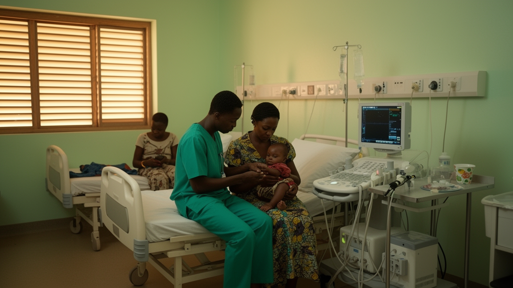
Image Credits: AI Generated
DOI: 10.1186/s12301-026-00622-1
Keywords: posterior urethral valves, paediatric urology, Africa, multicentre survey, delayed diagnosis, endoscopic valve ablation, chronic kidney disease, prenatal diagnosis, referral centres, healthcare resources, paediatric surgery, urinary tract obstruction
News Source: Ophelia Keating. (October 28, 2026). Survey of 31 African referral centres reveals delayed diagnosis and scarce endoscopic care for posterior urethral valves. Scienmag.