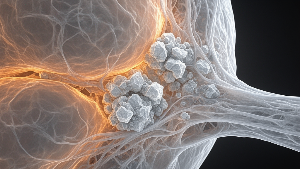
In clinical breast cancer screening, radiological evaluations have traditionally focused on the morphology and clustering patterns of microcalcifications, yet high-resolution three-dimensional imaging demonstrates that the primary diagnostic signal distinguishing benign from malignant lesions originates within the surrounding tissue interface rather than the calcified mineral core itself. In an ex vivo investigation evaluating ninety-four paraffin-embedded female breast biopsy specimens, researchers utilized three-dimensional micro-computed tomography at an isotropic resolution of approximately eight micrometers to isolate individual microcalcifications and their immediate tissue microenvironments. By systematically separating the internal mineral deposits from concentric surrounding tissue margins, the study demonstrated that machine learning classifiers trained exclusively on radiomic features from a narrow outer shell achieved diagnostic discriminative performance on par with models evaluating the entire expanded deposit and adjacent tissue. This finding directly challenges the conventional paradigm that treats mineralized specks as isolated diagnostic markers, establishing instead that microscopic tissue remodeling occurring within a band measuring only tens of micrometers around the calcification provides the dominant source of predictive quantitative imaging features.
Microcalcifications represent one of the earliest and most frequent signs of breast malignancy detected during routine mammographic screening, frequently triggering stereotactic core biopsies even when no palpable or visible soft-tissue mass is present. Approximately one-third of all malignant breast lesions are detected solely through the identification of these tiny mineral deposits, particularly in cases of ductal carcinoma in situ. In current clinical workflows, interpreting radiologists characterize microcalcifications using two-dimensional mammography and digital breast tomosynthesis according to Breast Imaging Reporting and Data System criteria, evaluating whether shapes appear coarse, round, fine pleomorphic, or linear branching. However, standard clinical imaging modalities operate with in-plane pixel dimensions ranging from fifty to one hundred micrometers and suffer from structural tissue superposition, masking microscopic boundary details. Although biological studies have indicated that active mineral deposition, stromal remodeling, and cellular interactions occur at the boundary between calcium deposits and adjacent tissue, conventional clinical equipment cannot resolve the internal architecture or the immediate surrounding tissue microenvironment of individual microcalcifications at single-digit micrometer scales.
To investigate where discriminative diagnostic information actually resides, the research team analyzed ninety-four archived stereotactic biopsy specimens obtained between 2007 and 2012 from female patients aged thirty-six to eighty-three years at Universitair Ziekenhuis Brussel. The cohort comprised forty-three histopathologically confirmed benign specimens and fifty-one malignant specimens, representing clinical presentations such as high-grade, micro-invasive, and low-grade ductal carcinoma in situ, lobular carcinoma in situ, fibrocystic changes, fibroadenomas, and hyperplasia. Formalin-fixed, paraffin-embedded tissue cores measuring approximately three millimeters in diameter were scanned on an ex vivo SkyScan 1076 micro-computed tomography system operated at sixty kilovolts and one hundred sixty-seven microamperes with an X-ray focal spot of approximately five micrometers. Each specimen underwent a twenty-four-minute projection sequence across one hundred eighty degrees of rotation, generating volumetric reconstructions with eight-micrometer voxels. A custom intensity-thresholding algorithm operating on an eight-bit scale segmented a total of 3,504 individual microcalcifications across the dataset, yielding 1,981 individual deposits from benign samples and 1,523 from malignant specimens, creating precise baseline three-dimensional masks for computational evaluation.
The experimental architecture evaluated handcrafted radiomic profiles across three sequential, tightly controlled computational investigations designed to isolate tissue signals from potential image processing artifacts. For each segmented microcalcification, investigators extracted up to 2,653 candidate radiomic features encompassing first-order voxel intensity statistics, three-dimensional shape metrics, and complex textural descriptors derived from Gray Level Co-occurrence, Run Length, Size Zone, Dependence, and Neighboring Gray Tone Difference matrices. These metrics were derived from raw grayscale data as well as mathematical image transformations, including Laplacian of Gaussian filtering and three-level Daubechies wavelet decompositions across eight directional sub-bands. To eliminate collinearity and reduce feature redundancy, pairwise Pearson correlation analysis discarded one feature from any pair exhibiting an absolute correlation above 0.95, halving overall dimensionality to between 800 and 1,500 features per case. Nine supervised machine learning algorithms, including Random Forest, Support Vector Machines, Multilayer Perceptrons, Extreme Learning Machines, and multiple gradient boosting variants, were trained under a leave-one-subject-out cross-validation framework incorporating internal nested validation and recursive feature elimination without clinical covariate inputs.
The initial experiment examined how the spatial extent of the rectangular image preprocessing window affects classification performance when the baseline calcification mask remains completely unchanged. In standard computational radiomics toolchains, operations like intensity normalization and filtering are executed across a cropped bounding box surrounding the region of interest prior to feature calculation, potentially permitting adjacent tissue grayscale values to alter voxels inside the segmentation mask. By incrementally widening the preprocessing window margin from zero up to thirty voxels beyond the calcification boundary, the researchers observed a marked increase in test-set area under the receiver operating characteristic curve, climbing from 0.748 at zero margin to 0.811 at a thirty-voxel expansion. The primary performance gains emerged in model sensitivity, which rose from 0.591 to 0.695, while model specificity remained comparatively steady between 0.817 and 0.851. Performance effectively reached an analytical plateau at a margin of ten voxels, demonstrating that unsegmented contextual tissue grayscale values significantly modulate features calculated within nominal segmentation boundaries.
To confirm that these performance improvements reflected true biological signal rather than technical preprocessing leakage, the team introduced a strict mask-only condition at the thirty-voxel window margin by setting all grayscale voxels outside the segmented core mask to zero prior to any filtering or extraction. When surrounding grayscale data were completely eliminated, model performance plummeted to an area under the curve of 0.688, with classification accuracy falling to 0.652 and the F1-score dropping to 0.573. Paired DeLong statistical tests with Bonferroni correction confirmed that every non-zero window margin significantly outperformed the zero-margin baseline and the masked-only configuration. Probabilistic calibration assessments further reinforced these findings, as the Brier score improved from 0.245 under zero margin down to approximately 0.167 across extended windows, well below the non-informative baseline of 0.25. The lowest expected calibration errors were achieved at intermediate window expansions between fifteen and twenty-five voxels, demonstrating that incorporating immediate peri-calcification context stabilizes both diagnostic discrimination and predictive probability reliability.
Building upon the preprocessing window findings, the second experimental phase evaluated the sensitivity of radiomic models to physical segmentation boundary shifts by applying morphological erosions and dilations ranging from two to ten voxels. Because each voxel represented eight micrometers, kernel adjustments of two, four, six, eight, and ten voxels corresponded to physical alterations of approximately sixteen, thirty-two, forty-eight, sixty-four, and eighty micrometers. Across all erosion and dilation steps, strict mask-only preprocessing was maintained by zeroing out voxels outside the modified region of interest. Progressively dilating the baseline mask into adjacent tissue produced a continuous, statistically significant rise in classification performance, with the area under the curve climbing from 0.689 at a two-voxel expansion to 0.813 at a ten-voxel dilation. Conversely, eroding the masks inward to capture only the dense internal calcification core yielded stagnant performance between 0.676 and 0.719, with paired DeLong tests showing no statistically significant differences relative to baseline after correction, confirming that restricting analysis to calcified cores discards essential diagnostic information.
The definitive third experiment isolated concentric volumetric shells to determine whether the surrounding microenvironment carries discriminative signal independently of the calcified core. Researchers extracted radiomic features from pure outer shells, defined by subtracting the baseline core mask from dilated volumes, and compared them directly against pure inner shells generated by subtracting eroded masks from the baseline. Outer shell classification models demonstrated a monotonic performance increase that tracked distance from the core, progressing from an area under the curve of 0.635 for the innermost two-voxel shell to 0.809 for the outermost ten-voxel shell. In stark contrast, inner shells derived strictly within the core boundary failed to surpass an area under the curve of 0.68. The best outer shell also achieved the lowest Brier score of 0.175 and an expected calibration error of 0.115, demonstrating that interface microtexture drives predictive accuracy.
Analysis of specific radiomic features across all testing conditions revealed that individual descriptors were highly sensitive to segmentation boundaries and surrounding tissue inclusion, though a robust subset consistently dominated high-performing classifiers. First-order distribution metrics capturing skewness and kurtosis alongside Gray Level Co-occurrence Matrix texture descriptors quantifying correlation and cluster prominence repeatedly demonstrated the highest discriminative power. Wavelet-transformed decompositions were frequently selected, indicating that multiscale gray-level heterogeneity carries more predictive utility than raw absolute intensity. Three-dimensional shape descriptors appeared inconsistently and failed to differentiate lesions reliably on their own. Biologically, the authors interpret these textural variations as imaging correlates of known histopathological phenomena, where malignant microenvironments foster disorganized mineralization, inflammatory infiltration, and active osteoblast-like stromal remodeling that generate marked gray-level heterogeneity, whereas benign microcalcifications exhibit homogeneous, stable interfaces. However, the authors caution that micro-computed tomography captures attenuation patterns rather than specific cellular or molecular pathways, meaning definitive biological mechanisms require future validation through co-registered histological staining.
The investigators emphasized several important methodological and clinical constraints governing the interpretation of their results. First, microcalcifications were labeled according to global specimen pathology, creating potential label noise because morphologically benign calcifications can reside within malignant biopsy specimens. Second, the study utilized archived, clinically suspicious stereotactic biopsy samples from a single institution without external scanner validation, reflecting an inherently suspicious cohort rather than a general screening demographic. Third, reconstructed volumes lacked calibration in standard Hounsfield units, restricting cross-study physical density comparisons, while the ex vivo setting and eight-micrometer resolution cannot be replicated directly on clinical mammography or digital breast tomosynthesis systems. Nevertheless, by demonstrating that microcalcification diagnostic information peaks within a narrow peri-calcification tissue band tens of micrometers wide, the authors conclude that future computational radiomics and computer-aided diagnosis frameworks must explicitly incorporate surrounding tissue interfaces to maximize diagnostic sensitivity and avoid discarding vital microenvironmental cues.
Subject of Research: Three-dimensional micro-CT radiomic analysis of breast microcalcification microenvironments
Article Title: Micro-CT radiomics reveals the microcalcification-surrounding tissue interface as the diagnostic epicenter of breast microcalcifications
Article References: Brahimetaj, R., Cornelis, J., Courtoy, G., Waelput, W., Willekens, I., De Mey, J., Forsyth, R., & Jansen, B. (2026). Micro-CT radiomics reveals the microcalcification-surrounding tissue interface as the diagnostic epicenter of breast microcalcifications. Breast Cancer Research, 28(1), Article 173. https://doi.org/10.1186/s13058-026-02362-0
Image Credits: AI Generated
DOI: 10.1186/s13058-026-02362-0
Keywords: micro-computed tomography, breast microcalcifications, radiomics, machine learning, tissue microenvironment, ductal carcinoma in situ, image preprocessing, biopsy specimens, receiver operating characteristic, feature extraction, concentric shells, digital breast tomosynthesis, diagnostic imaging, image segmentation, texture analysis, leave-one-subject-out
News Source: Ophelia Keating. (November 11, 2026). Surrounding Tissue Interface Dominates Breast Microcalcification Malignancy Predictions in Micro-CT Analysis. Scienmag.