A rare congenital venous anomaly that runs beneath the aortic arch could easily derail an operation for lung cancer, but surgeons in Japan have shown how careful preoperative imaging and a disciplined sequence of dissection can keep such an operation safe. In a case report published in General Thoracic and Cardiovascular Surgery Cases, a surgical team described removing the left upper lobe of the lung in a man in his seventies whose chest contained a subaortic anomalous left brachiocephalic vein, a vessel that crossed the aortopulmonary window and crowded the very space the surgeons needed to work in.
The vessel in question is an uncommon systemic venous anomaly in which the left brachiocephalic vein, instead of following its usual anterior route over the aortic arch, courses beneath it. In adult CT-based cohorts, the anomaly has been reported to be extremely rare, with incidences ranging from 0.03% to 0.062%. It is often associated with congenital heart disease or aortic arch anomalies, yet it is also detected incidentally in adults undergoing contrast-enhanced computed tomography for unrelated indications. For thoracic surgeons, its relevance becomes greatest when the anomalous vein traverses the aortopulmonary window, the narrow compartment between the aorta and the pulmonary artery, because it can restrict the operative corridor and raise the risk of vascular injury during left upper mediastinal dissection, including station 4L lymphadenectomy, as well as during exposure and control of the left pulmonary artery.
The patient at the center of the report was found to have a 4.2-centimeter solid mass in the left upper lobe, specifically in segment S3b. Computed tomography and fluorodeoxyglucose positron emission tomography showed enlarged lymph nodes at stations 4L and 11, along with bilateral lower-lobe reticulation and honeycombing consistent with a usual interstitial pneumonia pattern. No apparent FDG uptake was observed in the station 5 or 6 lymph node regions. Endobronchial ultrasound-guided transbronchial needle aspiration of the station 4L node demonstrated carcinoma consistent with non-small cell lung cancer.
Two factors shaped the treatment decision. The disease was judged to be resectable single-station N2 disease, meaning mediastinal nodal involvement confined to one lymph node station, but the patient also carried underlying interstitial lung disease with a usual interstitial pneumonia pattern. That fibrotic background raised concern for treatment-related pneumonitis, a feared complication in such patients, and after multidisciplinary discussion the team selected a surgery-first strategy with systematic lymphadenectomy. A preoperative biopsy of the primary lung lesion was not performed because the EBUS-TBNA had already confirmed mediastinal nodal malignancy, and definitive histology and molecular testing were planned using the resection specimen.
Before the operation, contrast-enhanced CT with three-dimensional reconstruction mapped the anomalous vessel in detail. The imaging demonstrated the left brachiocephalic vein coursing along the left lateral side of the aortic arch and then beneath the arch, superior to the pulmonary artery, and crossing the aortopulmonary window before draining into the superior vena cava. Based on these findings, the course was considered consistent with Takada pattern b/c, a classification that describes subaortic venous courses according to their relationship to the aortic arch, the ductus arteriosus ligament, and the pulmonary artery. The three-dimensional images showed the vein crossing the aortopulmonary window in close relation to the enlarged station 4L lymph node and the left pulmonary artery, allowing the surgical team to anticipate the restricted anatomy before making a single incision.
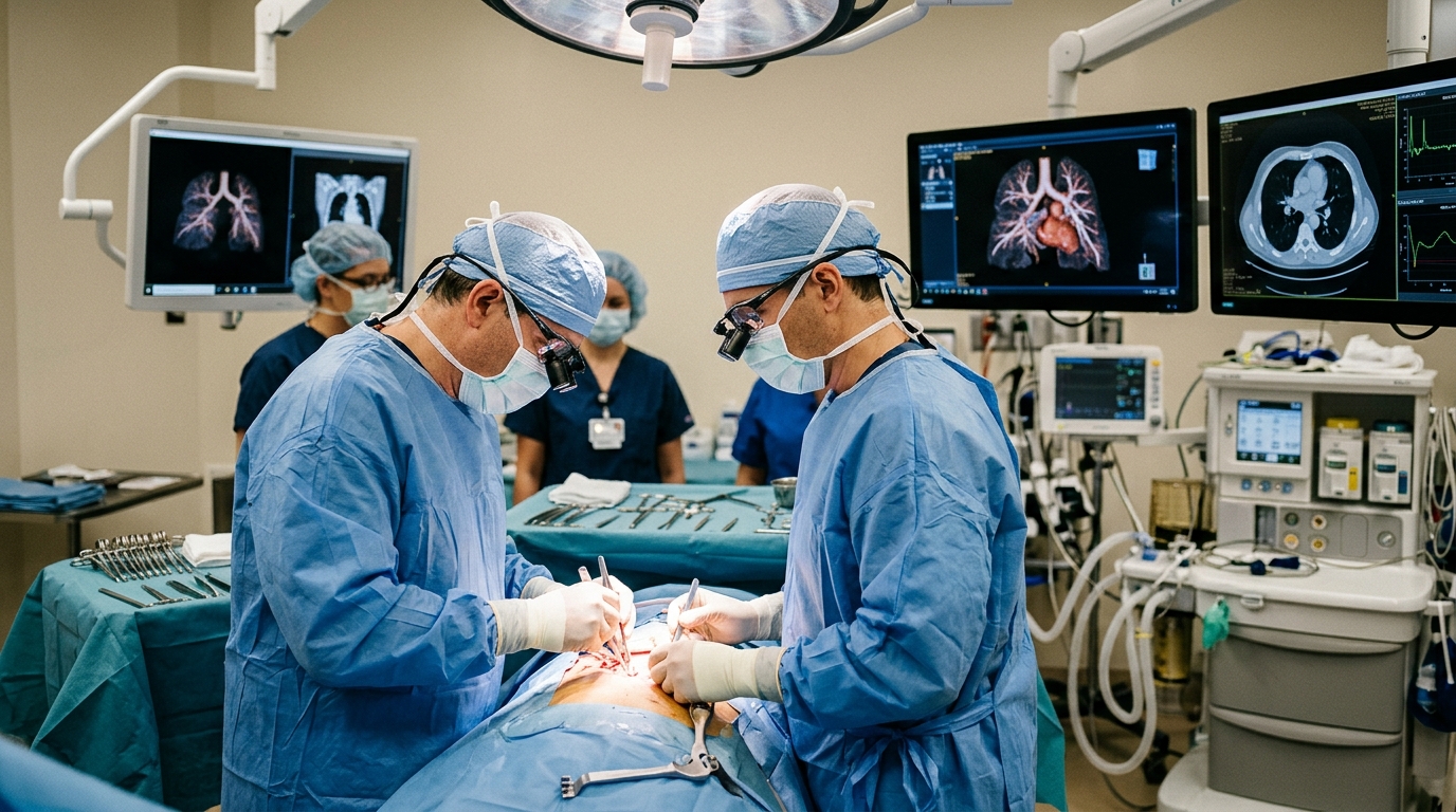
The operation itself was a video-assisted thoracoscopic left upper lobectomy with systematic mediastinal lymph node dissection. A 10-millimeter 30-degree thoracoscope was inserted through a camera port in the seventh intercostal space on the midaxillary line, and a 7-centimeter anterolateral utility thoracotomy was created through the fourth intercostal space. As predicted from the imaging, the subaortic anomalous vein occupied the aortopulmonary window, restricting the working space during station 4L lymphadenectomy and exposure of the left main pulmonary artery. The vessel passed beneath the aortic arch in close proximity to the station 4L and station 5 lymph node regions and the left pulmonary artery.
The operative strategy hinged on establishing reliable landmarks before deep dissection. The mediastinal pleura was opened on the dorsal mediastinal side of the hilum to identify the left vagus nerve, which was then dissected proximally to allow safe identification of the left recurrent laryngeal nerve. Although careful dissection was required because of the restricted operative field, identification of the recurrent laryngeal nerve itself was not technically difficult once the vagus nerve had been found. En bloc dissection of the station 4L and station 5 lymph node regions was performed after confirming the spatial relationship among the anomalous vein, the left pulmonary artery, the vagus nerve, and the recurrent laryngeal nerve. Pulmonary artery manipulation was deferred until a safe dissection plane had been established. No suspicious lymph node was identified in the station 6 region intraoperatively.
One anatomical detail remained unresolved. The ductus arteriosus ligament, the fibrous remnant of the fetal ductus arteriosus that anchors the classification system, could not be identified intraoperatively. Failure to find it in the expected location suggested a retroductal course, corresponding to Takada pattern c, in which the anomalous vein passes beneath the aortic arch and posterior to the ligament; however, definitive confirmation was not possible. The authors note that precise subtype classification may be difficult in adults because the ligament is not always identifiable on imaging or during surgery, and that in CT-based series, type b has been reported as the most common type according to Takada’s classification.
The operation was completed without vascular injury. The operative time was 2 hours and 29 minutes, and blood loss was 117 milliliters. The patient was discharged on postoperative day 4. Pathological examination revealed large cell carcinoma with mediastinal nodal metastasis, staged pT2bN2M0, stage IIIA according to the TNM 8th edition. Metastasis was identified in only one station 4L lymph node. The authors believe that the sequence they followed, early identification of the vagus and recurrent laryngeal nerves followed by careful pulmonary artery management after establishing a safe dissection plane, contributed to the safe completion of both the lymphadenectomy and the lobectomy without neural or vascular injury.
Only a limited number of reports have described lung resection in patients with a subaortic anomalous left brachiocephalic vein, and the authors suggest their case adds practical operative detail by showing how preoperative anatomical recognition and stepwise intraoperative dissection can help manage left-sided lung cancer when the anomalous vein constrains the aortopulmonary window. Previous radiologic series and review articles have emphasized the importance of recognizing this anomaly for operative planning and for avoiding misinterpretation as mediastinal lymphadenopathy, since a vessel crossing the window can be mistaken for abnormal tissue. The team’s conclusion is surgical rather than oncological: when a subaortic anomalous left brachiocephalic vein traverses the aortopulmonary window, safe surgery depends on accurate preoperative anatomical recognition with contrast-enhanced CT, with or without three-dimensional reconstruction, and disciplined intraoperative sequencing, with early identification of the vagus and recurrent laryngeal nerves providing reliable landmarks and pulmonary artery management undertaken only after a safe dissection plane has been established. This stepwise strategy may help prevent neural and vascular injury when left upper mediastinal dissection and pulmonary artery management are required within a constrained aortopulmonary window.
Subject of Research: A surgical case report describing left upper lobectomy for lung cancer in a patient with a rare subaortic anomalous left brachiocephalic vein traversing the aortopulmonary window.
Article Title: Left upper lobectomy for lung cancer with a subaortic anomalous left brachiocephalic vein: a case report
Article References: Sakata, R., Yoshida, Y., Yotsukura, M., Haruki, T., & Watanabe, S.-I. (2026). Left upper lobectomy for lung cancer with a subaortic anomalous left brachiocephalic vein: a case report. General Thoracic and Cardiovascular Surgery Cases, 5(1), Article 33. https://doi.org/10.1186/s44215-026-00266-1
Image Credits: AI Generated
DOI: 10.1186/s44215-026-00266-1
Keywords: lung cancer, left upper lobectomy, brachiocephalic vein, vascular anomaly, aortopulmonary window, thoracic surgery, VATS, lymph node dissection, three-dimensional CT, vagus nerve, recurrent laryngeal nerve, case report
News Source: Ophelia Keating. (November 5, 2026). Surgeons navigate rare vein anomaly to remove lung tumor safely. Scienmag.