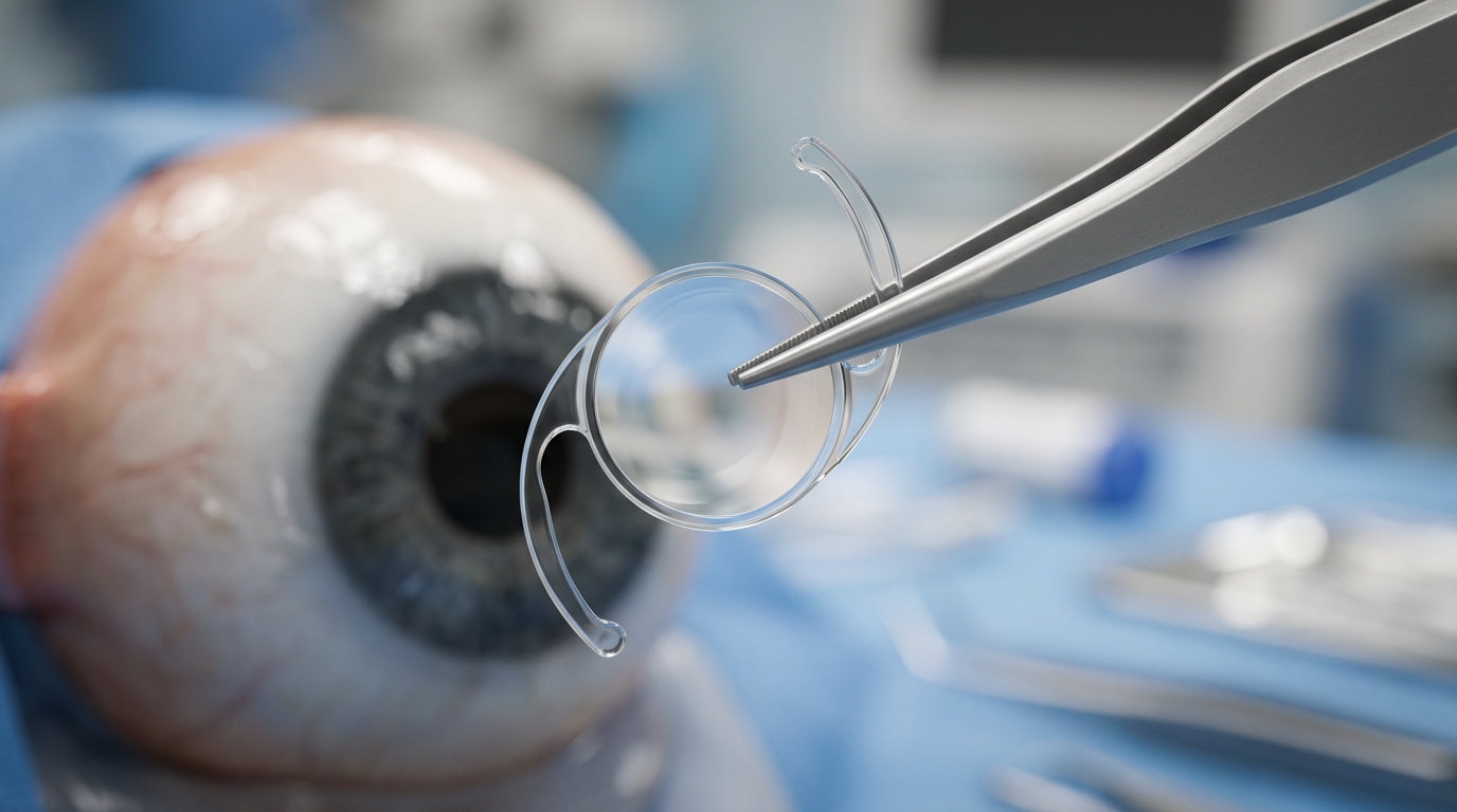
A retrospective longitudinal interventional case series conducted at the Hospital Regional Universitario de Málaga in Spain demonstrated that implanting supplemental intraocular lenses into the ciliary sulcus effectively reduced residual refractive errors and improved or stabilized distance visual acuity across sixteen eyes of eleven consecutive pseudophakic patients affected by severe, pre-existing ocular comorbidities. The surgical intervention utilized the AddOn supplemental intraocular lens platform manufactured by 1stQ Deutschland GmbH in both spherical and toric designs. Throughout a mean postoperative follow-up duration of 25.75 ± 7.57 months, ranging from 12 to 39 months, every treated eye experienced a measurable reduction in postoperative residual refractive error, while monocular uncorrected distance visual acuity improved in the majority of treated eyes. Importantly, monocular corrected distance visual acuity remained entirely stable or improved across all sixteen eyes, with no patient losing any lines of best-corrected visual acuity. Furthermore, the clinical cohort experienced no intraoperative complications and zero postoperative adverse events, presenting no instances of elevated intraocular pressure, pupillary block, pigment dispersion, chronic intraocular inflammation, or corneal decompensation over the extended monitoring period.
Standard cataract surgery remains one of the most widely performed and cost-effective surgical procedures worldwide, typically delivering predictable and highly satisfactory visual restoration. However, achieving precise target refraction becomes exceptionally difficult in eyes characterized by complex anatomical anomalies, extreme axial dimensions, or coexisting corneal and retinal pathologies. Postoperative pseudophakic refractive surprises frequently arise from keratometric inaccuracies in irregular corneas, post-refractive ectasia, or prior corneal grafts, as well as biometry errors in eyes exhibiting extreme axial lengths, instrumentation discrepancies, human calculation mistakes, or rare manufacturer lens mislabelling. Historically, managing these debilitating refractive errors required intraocular lens explantation and exchange, a technically demanding and hazardous intraocular maneuver carrying heightened risks of capsular rupture, zonular dialysis, corneal endothelial trauma, and retinal detachment. While corneal laser enhancement offers an alternative corrective avenue, it is frequently contraindicated in thin, irregular, or transplanted corneas. In such challenging clinical scenarios, placing a secondary supplemental intraocular lens directly into the ciliary sulcus in front of the preserved, intact capsular bag provides a tissue-sparing, reversible solution that bypasses the substantial surgical hazards associated with primary lens removal or corneal ablation.
The study cohort comprised eleven consecutive patients representing sixteen individual eyes treated between routine clinical appointments by a single experienced ophthalmic surgeon. The patient population exhibited a mean age of 63.27 ± 16.43 years, with ages spanning widely from 21 to 84 years, reflecting the diverse origins of complex ocular pathology encountered in tertiary clinical care. Eligible participants were pseudophakic individuals with an intact, stable capsular bag containing a primary intraocular lens alongside documented anterior or posterior segment comorbidities who completed a minimum of 12 months of structured postoperative evaluation. Patients who lacked sufficient clinical records, had shorter follow-up durations, or underwent unrelated secondary ocular procedures that could confound visual outcomes were excluded from the analysis. The pre-existing comorbidities encompassed an extraordinarily wide spectrum of challenging conditions, including keratoconus, post-laser-assisted in situ keratomileusis ectasia, prior penetrating keratoplasty, extreme axial myopia, radial and transverse incisional keratotomies, advanced glaucoma, atrophic age-related macular degeneration, inactive ocular toxoplasmosis, previous surgical retinal detachment repair, severe ocular surface dry eye disease, and corneal dystrophies.
Surgeons executed the clinical protocol under strict procedural parameters, postponing supplemental lens selection until at least three months had elapsed following primary cataract surgery. This mandatory observation interval ensured that corneal biomechanics and capsular healing had achieved complete refractive stability, confirmed through consistent serial manifest subjective refractions without spontaneous fluctuation. Unlike primary intraocular lens formulas that rely heavily on complex anatomical anterior chamber depth assumptions and corneal curvature models, the calculation for the AddOn supplemental lens relies primarily on the patient’s manifest subjective refraction yielding maximal corrected distance visual acuity, supplemented by anatomical biometry data. Preoperative biometry was gathered using the IOLMaster 500 optical biometer operated in pseudophakia mode alongside Pentacam HR Scheimpflug corneal tomography. The subjective manifest refraction was inputted into the manufacturer’s proprietary web-based calculator. Toric supplemental intraocular lenses were deliberately selected for eyes demonstrating clinically significant subjective improvement with astigmatic corrections of approximately 1.50 diopters or greater, while spherical designs addressed purely spherical refractive surprises. All eyes were targeted for postoperative emmetropia, with the deliberate exception of one patient targeted for myopic undercorrection.
Surgical implantation of the AddOn supplemental intraocular lenses followed an established microincisional technique performed under sterile conditions without prophylactic iridotomy or iridectomy. The surgeon created a 15-degree auxiliary paracentesis followed by a 2.75-millimeter primary clear corneal incision. For toric lens models, the target alignment meridian was precisely marked on the cornea prior to entry. The anterior chamber was maintained and stabilized using an injection of Fydrane ophthalmic solution alongside a high-molecular-weight cohesive ophthalmic viscosurgical device comprising sodium hyaluronate. The foldable supplemental intraocular lens was injected and manipulated directly into the ciliary sulcus, positioned carefully over the intact anterior capsule without disturbing the primary lens seated firmly within the capsular bag, and rotated into proper alignment when toric optics were utilized. Following thorough aspiration and complete evacuation of the viscosurgical device from the anterior segment, the surgeon administered intracameral cefuroxime for infection prophylaxis and intracameral acetylcholine to induce miosis, concluding the intervention with balanced salt solution stromal hydration of both incisions to achieve watertight closure.
Detailed postoperative evaluations conducted at six months and at the final follow-up visit confirmed substantial refractive and optical improvements across the cohort. Postoperative spherical equivalent measurements shifted markedly closer to emmetropia in the vast majority of treated eyes. Monocular uncorrected distance visual acuity improved in most treated eyes, while every eye maintained or improved its baseline corrected distance visual acuity, demonstrating that the secondary optical interface produced no loss of visual quality. Individual cases illustrated the practical versatility of this approach across complex scenarios. For instance, patient number 8, who experienced a substantial refractive surprise attributable to a primary biometry calculation error, attained complete postoperative emmetropia alongside marked gains in uncorrected visual acuity. Similarly, patient number 10, affected by severe dry eye disease and high irregular astigmatism, experienced successful refractive neutralization and enhanced visual function without surface compromise. Across all sixteen surgical procedures, no secondary surgical interventions, such as intraocular lens repositioning, rotational realignment, or explantation, were required during the monitoring timeline.
The study also highlighted several particularly intricate cases managed with planned sequential or rescue strategies. One representative patient presented with extreme axial myopia measuring 32.93 millimeters in the right eye and 34.36 millimeters in the left eye, complicated by prior bilateral anterior chamber Cachet phakic lenses that caused pupillary miosis and extensive posterior synechiae. Because optical biometry was impossible, ultrasound biometry was required, further compounded by previous radial keratotomy, transverse keratotomy, and corneal laser enhancement. The surgical team executed a pre-planned two-stage approach, initially performing cataract extraction with phakic lens explantation through a scleral tunnel, followed months later by supplemental lens implantation to correct the residual refractive error of minus 8.00 diopters sphere with minus 3.25 diopters cylinder in the right eye and minus 4.00 diopters sphere with minus 3.00 diopters cylinder in the left eye. Another challenging case involved post-refractive ectasia with extreme corneal thinning that strictly prohibited intracorneal ring segments, where an AddOn spherical lens successfully corrected a postoperative surprise of plus 5.50 diopters sphere with minus 1.25 diopters cylinder. Similarly, a patient with a synechiated mature cataract following penetrating keratoplasty successfully received a toric supplemental lens after stabilizing.
Beyond standard clinical visual acuity metrics, the study evaluated subjective patient-reported outcome measures by administering validated instruments to eight of the eleven patients who attended an additional research visit. Functional visual performance was measured using the 14-item Visual Function Index, with scores spanning from 23.21 to 100.00 across the cohort, mirroring the substantial diversity of underlying ocular comorbidities. Five of the eight surveyed individuals achieved high functional scores, including two patients achieving perfect scores of 100.00 and three scoring 91.07, 88.64, and 76.79. The single patient recording the lowest functional score of 23.21 suffered from advanced bilateral atrophic age-related macular degeneration, presenting identical uncorrected and corrected distance visual acuities of 0.40 logMAR despite near-emmetropic postoperative refraction of minus 0.25 diopters sphere with minus 0.75 diopters cylinder at 80 degrees, confirming that systemic retinal degeneration governed the functional limitation rather than residual optical error.
Analysis of the Patient-Reported Spectacle Independence Questionnaire revealed high rates of functional independence, with six of the eight patients, or 75 percent, achieving complete freedom from spectacles for distance viewing tasks. Although uncorrected intermediate and near visual rehabilitation was not the primary therapeutic objective of these monofocal and toric implants, six patients also reported complete independence from intermediate spectacles, while two patients attained near spectacle independence. Qualitative survey questions revealed that seven of eight patients experienced positive satisfaction with their distance vision, and all eight participants reported satisfaction with intermediate vision. When questioned regarding positive dysphotopsia, six patients noted only slightly bothersome photic phenomena, two patients reported no bothersome phenomena whatsoever, and zero patients reported moderate or severe dysphotopsia. Crucially, when asked whether they would choose the procedure again, 100 percent of respondents confirmed that they would willingly undergo the supplemental lens implantation.
The study authors acknowledged significant methodological limitations inherent to an uncontrolled retrospective case series involving a modest sample size of sixteen eyes across eleven patients. The reliance on existing clinical charts carried inherent risks of selection bias, and the absence of a parallel control cohort undergoing alternative interventions precluded comparative efficacy or causal assertions. Furthermore, combining planned two-stage sequential procedures with rescue treatments for unexpected refractive surprises pooled disparate clinical indications. The investigators also noted that objective rotational alignment of the toric supplemental lenses was not systematically quantified using imaging analysis, endothelial cell densities were not longitudinally tracked with specular microscopy, and comprehensive formal health economics and device cost data were not available. Nevertheless, the prolonged follow-up period confirmed the clinical feasibility, safety, and reversibility of sulcus-based AddOn supplemental intraocular lenses, demonstrating their utility when corneal laser surgery or primary lens explantation carries unacceptable ocular risks.
Subject of Research: Supplemental intraocular lens implantation for residual refractive error in eyes with complex ocular comorbidities
Article Title: Visual and Patient-Reported Outcomes Following Supplemental Intraocular Lens Implantation for Pseudophakic Refractive Error in Eyes with Complex Ocular Comorbidities
Article References: Rocha-de-Lossada, C., Burguera, N., Estévez Domínguez, R., Rodríguez-Vallejo, M., Calvo Rodríguez-de-Mora, M., & Fernández, J. (2026). Visual and Patient-Reported Outcomes Following Supplemental Intraocular Lens Implantation for Pseudophakic Refractive Error in Eyes with Complex Ocular Comorbidities. Ophthalmology and Therapy. https://doi.org/10.1007/s40123-026-01518-7
Image Credits: AI Generated
DOI: 10.1007/s40123-026-01518-7
Keywords: ophthalmology, supplemental intraocular lens, pseudophakic refractive error, AddOn lens, cataract surgery, ocular comorbidities, sulcus implantation, keratoconus, penetrating keratoplasty, visual acuity, patient-reported outcomes, spectacle independence, refractive surgery, astigmatism, toric intraocular lens, retrospective case series
News Source: Ophelia Keating. (November 17, 2026). Supplemental Intraocular Lenses Correct Pseudophakic Refractive Errors in Complex Eyes. Scienmag.