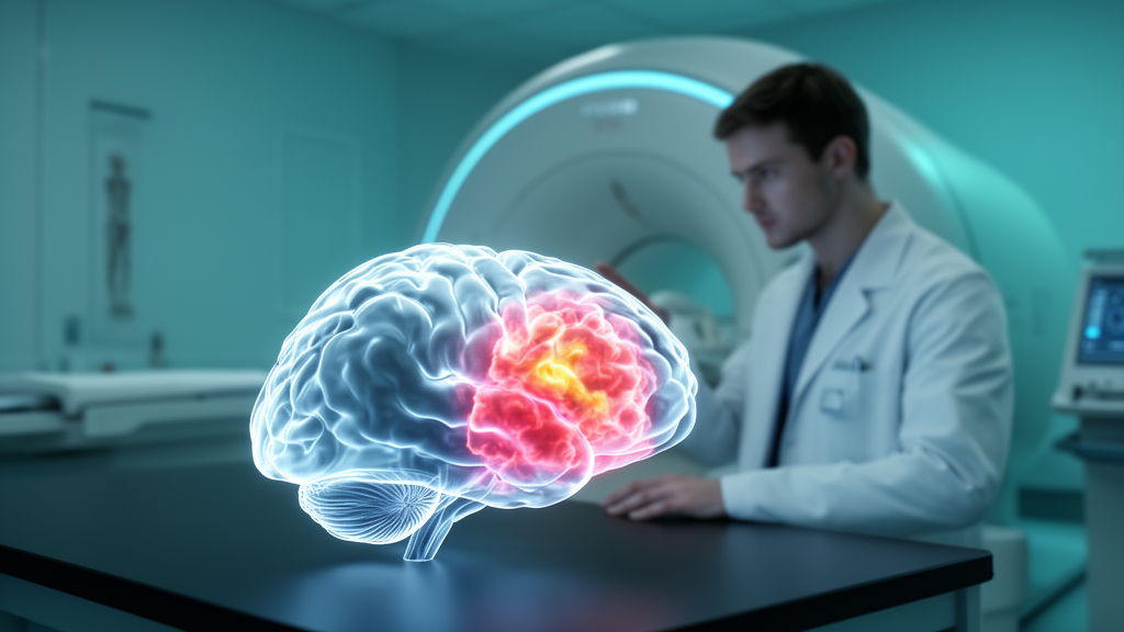
A quantitative magnetic resonance imaging technique that can separate paramagnetic from diamagnetic substances within a single imaging voxel has revealed distinct magnetic susceptibility profiles across different regions of brain tumors, according to a new study published in European Radiology Experimental. The research, led by Shanmei Zeng, Nengjin Zhu and Junqiao Wang with colleagues at the First Affiliated Hospital of Sun Yat-sen University, applied the sub-voxel quantitative susceptibility mapping method known as APART-QSM to 120 patients with pathologically confirmed gliomas, examining tumor parenchyma, peritumoral edema, necrosis and cystic degeneration as separate compartments rather than treating the tumor as a single mass.
Conventional quantitative susceptibility mapping measures a voxel-averaged susceptibility value, which cannot distinguish between paramagnetic and diamagnetic components coexisting within the same voxel. In gliomas, where complex tissue constituents frequently overlap spatially, this averaging can produce signal cancellation effects that obscure the true susceptibility profile. APART-QSM addresses this limitation by employing iterative data fitting to simultaneously separate and quantify paramagnetic and diamagnetic substances within a voxel, such as iron and myelination. The technique has previously been applied to neurological disorders including attention-deficit/hyperactivity disorder, epilepsy and Alzheimer’s disease, but its potential for comprehensive glioma characterization had remained largely unexplored before this study.
The retrospective cohort comprised 120 patients aged 47 ± 13 years, of whom 51 were women, drawn from an initial screen of 179 patients who underwent preoperative MRI between February 2019 and October 2023. All tumors were pathologically confirmed, and surgical resection was performed within two weeks of the MRI examination. The cohort included 59 patients with isocitrate dehydrogenase (IDH)-mutant gliomas and 61 with IDH-wildtype gliomas, with 1p/19q co-deletion present in 20 IDH-mutant and one IDH-wildtype case. Imaging was performed on a 3-Tesla scanner using a three-dimensional multi-echo gradient-echo sequence acquired before contrast administration, and the APART-QSM post-processing pipeline included brain extraction, phase unwrapping, V-SHARP, STAR-QSM and iterative separation steps.
Regions of interest were manually delineated in consensus by two experienced neuroradiologists, with a third blinded neuroradiologist independently delineating the same regions to assess reproducibility. Interobserver agreement was excellent across all tumor subregions. To analyze susceptibility changes in peritumoral edema at increasing distances from the tumor, the researchers progressively dilated the tumor mask outward and divided the total edema region into four equidistant concentric subregions, labeled Edema1 through Edema4 from nearest to farthest. Contralateral normal-appearing tissue masks were generated by mirroring the combined tumor and edema mask across the midline, with overlapping lesion, ventricular and extra-cranial areas excluded.
The results showed clear contrasts between tumor subregions and between tumor subtypes. In high-grade gliomas, tumor parenchyma exhibited significantly higher paramagnetic susceptibility than contralateral normal-appearing tissue (p = 0.044), while low-grade gliomas showed significantly lower bulk, paramagnetic and diamagnetic susceptibility (p ≤ 0.006). IDH-mutant gliomas displayed significantly lower bulk, paramagnetic and diamagnetic susceptibility in tumor parenchyma than contralateral tissue (p ≤ 0.015), whereas IDH-wildtype gliomas showed significantly elevated diamagnetic and paramagnetic susceptibility (p ≤ 0.034). High-grade tumors and IDH-wildtype tumors each showed significantly higher paramagnetic and diamagnetic susceptibility in tumor parenchyma than their lower-grade or IDH-mutant counterparts (p < 0.001).
Peritumoral edema showed significantly lower bulk and paramagnetic susceptibility than contralateral normal-appearing tissue in all gliomas (p < 0.001), a pattern the authors attribute to the elevated water content of edema, which dilutes susceptibility signals. Notably, within the edema subregions, both paramagnetic and diamagnetic susceptibility followed a non-linear spatial pattern, initially decreasing and then increasing with distance from the tumor margin (p ≤ 0.008). Paramagnetic susceptibility was highest in the nearest subregion and lowest in the second subregion relative to all others, with differences reaching statistical significance under Bonferroni correction. The authors suggest this distance-associated pattern may reflect the dynamic interaction between infiltrating iron-rich tumor cells and inflammatory cells and the degree of edema, which contains abundant weakly diamagnetic water.
Necrotic regions exhibited elevated bulk, diamagnetic and paramagnetic susceptibility compared with contralateral tissue (p ≤ 0.025), with high-grade gliomas and IDH-wildtype gliomas showing significant increases in necrosis. The authors propose that elevated paramagnetic content in necrosis may arise from thrombosis, hypoxia, blood-brain barrier disruption and neovascularization, while elevated diamagnetic susceptibility may reflect necrotic debris and denatured proteins, with underlying mechanisms requiring further elucidation. Cystic degeneration showed no significant susceptibility differences overall, but in oligodendrogliomas, cystic degeneration displayed lower bulk susceptibility yet higher diamagnetic susceptibility relative to contralateral tissue, a pattern potentially attributable to intratumoral calcification, whereas non-oligodendrogliomas showed lower diamagnetic susceptibility, consistent with a predominance of weakly diamagnetic water.
To explore whether multi-subregional susceptibility profiles could classify gliomas by molecular genotype, the researchers built a conditional inference tree model using twelve susceptibility predictors to distinguish three groups: IDH-mutant tumors without 1p/19q co-deletion, IDH-mutant tumors with co-deletion, and IDH-wildtype tumors. The model identified diamagnetic susceptibility in tumor parenchyma and paramagnetic susceptibility in peritumoral edema as the primary predictors, achieving an accuracy of 0.767 in the training set and 0.702 in the testing set. However, performance was considerably limited for the co-deleted subtype, with zero sensitivity in both sets, a limitation the authors attribute to data imbalance in the cohort, given the limited number of oligodendroglioma cases. They emphasize that the model is exploratory and requires validation in larger, more balanced cohorts before any clinical implementation.
Survival analysis revealed that higher paramagnetic and diamagnetic susceptibility in tumor parenchyma were significantly associated with shorter overall survival in univariable analysis (both p < 0.001 by log-rank test), with hazard ratios of 1.021 and 1.037 respectively. However, in multivariate Cox regression, only age remained an independent prognostic factor, suggesting that the susceptibility associations may be confounded by other clinical factors. In a prospectively enrolled subgroup of six patients, the researchers also validated the pathological basis of the measurements: paramagnetic susceptibility in tumor parenchyma correlated strongly with histological iron content measured by Perls’ Prussian blue staining (r = 0.901, p = 0.014), and both paramagnetic and diamagnetic susceptibility correlated with the Ki-67 proliferation index (r = 0.841, p = 0.036). The authors caution that these preliminary findings warrant validation in larger cohorts.
The study carries several important limitations that the authors themselves acknowledge. Most subgroup analyses used nominal, uncorrected p-values and should be interpreted as exploratory. The sample size was modest, particularly for subgroups including oligodendrogliomas, tumors with cystic degeneration or necrosis, and the six-patient histologic validation cohort. The conditional inference tree model lacked an external validation cohort, and the exclusion of technically suboptimal cases may have introduced selection bias. The relationship between lipid accumulation, calcification and diamagnetic susceptibility was not directly validated, and no pathological confirmation was obtained for the susceptibility changes observed in peritumoral edema subregions or necrosis. Despite these constraints, the study demonstrates that sub-voxel susceptibility mapping can quantify distinct magnetic susceptibility profiles across glioma subregions, offering quantitative imaging biomarkers for iron content and tumor proliferation and a suggestive, though not independent, association with patient prognosis, with the peritumoral edema pattern potentially reflecting microenvironmental properties that merit further investigation.
Subject of Research: Sub-voxel quantitative susceptibility mapping of magnetic susceptibility patterns across glioma subregions and their clinicopathological correlations
Article Title: Multi-subregional susceptibility patterns in gliomas using sub-voxel quantitative susceptibility mapping
Article References: Zeng, S., Zhu, N., Wang, J., Mazu, L., Huang, Y., Xia, Z., He, S., Wu, J., Qin, H., Yang, Z., Liu, X., Wei, H., & Chu, J. (2026). Multi-subregional susceptibility patterns in gliomas using sub-voxel quantitative susceptibility mapping. European Radiology Experimental, 10(1), Article 164. https://doi.org/10.1186/s41747-026-00847-3
Image Credits: AI Generated
DOI: 10.1186/s41747-026-00847-3
Keywords: glioma, quantitative susceptibility mapping, APART-QSM, MRI, peritumoral edema, IDH mutation, iron deposition, Ki-67, tumor necrosis, cystic degeneration, genotyping, prognosis
News Source: Nathaniel Bowman. (October 28, 2026). Sub-Voxel MRI Technique Maps Distinct Magnetic Susceptibility Patterns Across Glioma Subregions. Scienmag.