The escalating prescription of GLP-1 receptor agonists, medicines primarily targeting type 2 diabetes and weight management, is presenting an emerging challenge in the realm of oncological imaging—specifically concerning the interpretation of FDG PET-CT scans. New research unveiled at the 38th Annual Congress of the European Association of Nuclear Medicine (EANM’25) in Barcelona has highlighted the complexity that these drugs introduce into metabolic imaging, signaling a need for revised evaluation protocols to avoid diagnostic pitfalls.
GLP-1 receptor agonists have revolutionized metabolic disorder management, exhibiting exponential growth in usage; in the United States alone, patient numbers without diabetes prescribed these drugs surged by an astonishing 700% from 2019 to 2023. Their multifaceted pharmacological effects, including modulation of glucose metabolism, slowing gastric emptying, and altering sympathetic nervous system activity, directly influence the biodistribution patterns of radiotracers such as fluorodeoxyglucose (FDG) used in PET-CT scans. These physiological alterations yield atypical patterns of tracer uptake, complicating the clear differentiation between malignant versus benign or inflammatory processes.
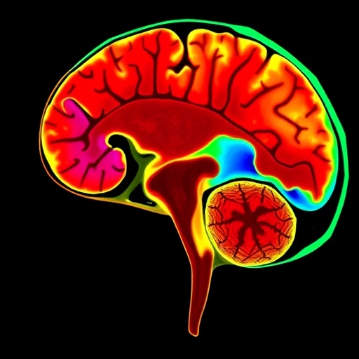
Historically, FDG PET-CT has been a cornerstone in oncological diagnostics, capitalizing on the increased glycolytic activity of cancer cells to localize malignant tissues. However, the novel uptake signatures observed in patients on GLP-1 agonists—even in non-malignant tissues like skeletal muscle, myocardium, and brown adipose tissue—raise the specter of false-positive results, potentially mimicking aggressive neoplastic or inflammatory pathologies. Earlier isolated case reports have documented such misleading uptakes, but the current study is the first to systematically characterize these phenomena, underscoring a critical gap in interpretative clarity.
Dr. Peter Strouhal, Medical Director at Alliance Medical Ltd, led an extensive retrospective analysis focusing on oncological FDG PET-CT scans from patients receiving GLP-1 analogue therapy. This large-scale review exposed a spectrum of altered tracer distribution patterns that if unrecognized, harbored profound implications for clinical decision-making. The research team observed consistent, reproducible uptake anomalies that diverged from classic oncological imaging phenotypes, necessitating a nuanced approach that incorporates comprehensive medication histories.
The implications of misread scans are far-reaching. While false positives catalyze unnecessary diagnostic interventions, biopsies, and even inappropriate staging, they also impose psychological distress and inevitable delays in therapeutic timelines for patients. Dr. Strouhal emphasized the clinical imperative to recognize these distinctive imaging effects—not to alarm clinicians but to enhance diagnostic precision and patient care pathways. The avoidance of unwarranted anxiety and intervention is paramount to optimizing resource utilization and upholding the integrity of oncologic management.
Currently, no formalized international or UK-specific guidance exists to address these GLP-1 agonist-induced PET-CT interpretation challenges. Though preliminary recommendations from Australian entities like the joint ADS/ANZSNM guidelines offer some direction—advocating for continuation of therapy, overnight fasting, morning scan scheduling, and stringent glycemic control—these remain regional suggestions without broad consensus. This regulatory vacuum underscores the urgency for coordinated efforts to adapt imaging protocols worldwide.
Rather than recommending cessation of GLP-1 receptor agonists prior to FDG PET-CT, which might disrupt metabolic homeostasis or compromise chronic disease control, experts suggest meticulous documentation of patient pharmacotherapy in imaging requisitions and reports. Detailed medication histories equip nuclear medicine specialists to contextualize scan findings accurately, integrating clinical pharmacology with metabolic imaging biomarkers to distinguish therapeutic effects from true pathologic activity.
Beyond immediate clinical practice, Alliance Medical Ltd’s team plans to expand their research network, pooling data from multiple imaging centers to build a robust evidence base that can drive national guidelines. They also envision forming international collaborations aimed at harmonizing protocols and enhancing global PET-CT interpretative consistency, a crucial step toward safeguarding diagnostic certainty as GLP-1 agonist usage proliferates worldwide.
Technologically, these findings illustrate the dynamic interface between emerging therapeutics and imaging science, spotlighting the necessity for continuous updates to nuclear medicine interpretation frameworks. Notably, the FDG tracer, a glucose analogue, is exquisitely sensitive to metabolic perturbations; thus, therapies that recalibrate systemic glucose handling inherently interfere with its uptake patterns. Understanding these pharmacodynamics is essential for molecular imaging specialists to maintain diagnostic accuracy.
The research also invites further mechanistic exploration into how GLP-1 receptor activation modulates physiological glucose uptake pathways across diverse tissues. Identifying the precise biochemical and cellular bases for these aberrant FDG signal amplifications could unlock biomarkers for therapy monitoring, expanding the diagnostic utility of PET imaging beyond oncology to metabolic therapeutic surveillance.
In conclusion, the remarkable rise of GLP-1 receptor agonists heralds a new era in metabolic medicine but concomitantly challenges oncological imaging paradigms. This pioneering research from EANM’25 not only cautions against diagnostic misinterpretations in FDG PET-CT scans but propels the nuclear medicine community to innovate adaptive guidelines and heighten interdisciplinary awareness. By integrating pharmacotherapeutic context into imaging workflows, healthcare teams can ensure that patients receive accurate, timely cancer diagnoses without unnecessary detours or distress, ultimately advancing precision medicine.
Subject of Research: Effects of GLP-1 receptor agonists on FDG PET-CT imaging interpretation in oncology.
Article Title: GLP-1 Receptor Agonists and Their Complex Impact on Oncological FDG PET-CT Scan Accuracy: Insights from the 38th Annual EANM Congress.
News Publication Date: Wednesday, 8 September 2025.
Web References: For further inquiries or expert interviews, contact [email protected].
References:
Strouhal P, Meadows A, McGovern A. A Weighty Problem: GLP-1 Agonists and the Altered Images of FDG PET-CT. Presented at EANM’25, 8 October 2025.
Mahase E. GLP-1 agonists: US sees 700% increase over four years among non-diabetic patients. BMJ, 2024.
Oldan JD, Landman PG, Schroeder JA et al. FDG PET in a Patient on a GLP-1 Agonist/Insulin Secretagogue. Clinical Nuclear Medicine, 2024.
Harrison DB, Phillips AL, Tansey JB et al. Brown Adipose Tissue Mimicking Head and Neck Cancer on PET Scan in a Patient on GLP-1 Drug. Laryngoscope, 2025.
Greenfield J, Mikaheal Y, Ludington J. Joint ADS/ANZSNM guideline for FDG PET/CT imaging in patients with type 1 and type 2 diabetes. Australian Diabetes Society & ANZSNM, 2024.
Keywords: GLP-1 receptor agonists, FDG PET-CT, oncology imaging, metabolic disorders, diabetes, weight management, diagnostic imaging, molecular imaging, nuclear medicine, pharmacotherapy impact, cancer staging, imaging artifacts.
Tags: cancer detection challengesdiagnostic pitfalls in imagingfluorodeoxyglucose uptake patternsGLP-1 receptor agonistsmetabolic disorder treatment innovationsmetabolic imaging complexitiesoncological diagnosticsPET-CT imaging challengesphysiological effects of GLP-1 agonistsprescription growth in diabetes drugsrevised evaluation protocolstype 2 diabetes medications

