In a groundbreaking advancement in neuropharmacology, researchers have unveiled detailed structural snapshots revealing the elusive process of nucleotide release at the μ-opioid receptor (MOR). This discovery provides unprecedented insight into the molecular mechanisms underlying opioid receptor signaling, a critical pathway influencing pain management and addiction. Utilizing cutting-edge cryo-electron microscopy (cryoEM) and sophisticated biochemical assays, the team has elucidated how MOR interacts with G proteins and nucleotides during activation and inhibition, paving the way for next-generation therapeutics with improved safety profiles.
The μ-opioid receptor is a G protein-coupled receptor (GPCR) that mediates the effects of opioid drugs, which are among the most potent analgesics but also notorious for their addictive potential. Despite decades of research, the dynamic conformational changes and nucleotide exchange events within MOR-G protein complexes have remained poorly understood. This study bridges that knowledge gap by capturing the receptor in various functional states, including inactive, GDP-bound, nucleotide-free, and GDP-rebound conformations, through meticulous structural and functional characterization.
Expression and purification of MOR and associated proteins posed significant challenges due to their membrane-embedded nature and conformational flexibility. The team employed recombinant expression systems leveraging insect cells (Spodoptera frugiperda) and human embryonic kidney cells to obtain high yields of functional receptor protein. Advanced affinity purification strategies, including tandem His and Flag tags, enabled isolation of pure receptor complexes suitable for high-resolution structural studies.
To characterize receptor conformations, researchers utilized nuclear bathrobe-like nanobody Nb6M and heterotrimeric G protein subunits co-expressed with MOR. These complexes were stabilized with ligands such as naloxone and loperamide, known antagonists and agonists, respectively, to mimic distinct physiological states. The meticulous preparation ensured the preservation of native-like receptor conformations critical for downstream cryoEM and biochemical assays.
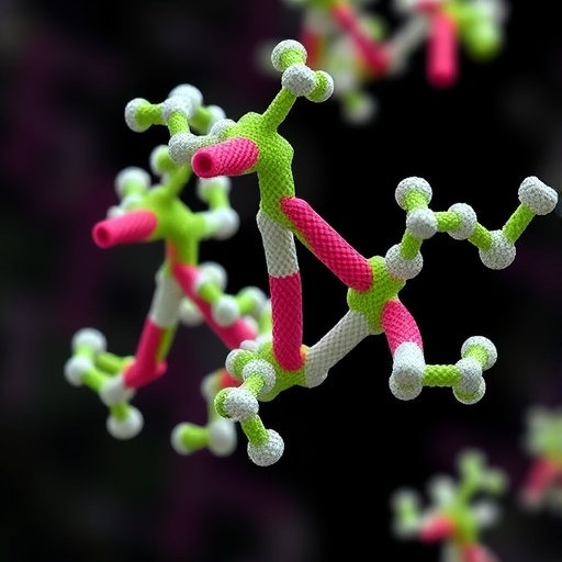
Cutting-edge cryo-electron microscopy allowed visualization of the MOR-G protein interface at near-atomic resolution, revealing subtle but critical movements within the receptor and G protein heterotrimer upon nucleotide release. Advanced single-particle reconstruction techniques led to maps resolving key regions involved in signal transduction, such as the transmembrane domain (TMD) and the α-helical domain (AHD) of Gα subunits. These snapshots captured transient states previously inaccessible to structural biology.
In parallel, bioluminescence resonance energy transfer (BRET) assays were employed to monitor real-time interactions and competition events between MOR and G protein subunits in living cells. These sensitive assays quantified nucleotide binding affinities and the effect of various ligands on receptor activation dynamics. The data revealed distinct ligand-specific modulations in nucleotide exchange rates, further correlating structural states with functional outcomes.
Complementing the structural and biophysical approaches, radioligand saturation binding experiments quantified the affinity of ligands toward MOR in membrane preparations, confirming the functional relevance of purified constructs. These experiments established the competitive binding profile of naloxone, loperamide, and other compounds in the presence of radiolabeled naltrexone, ensuring the biological validity of the receptor complexes studied.
The article also highlights the deployment of molecular dynamics (MD) simulations that provided atomistic insights into receptor-ligand and receptor-G protein interactions over microsecond timescales. By embedding MOR-G protein complexes within realistic lipid bilayer environments, the simulations captured energetic landscapes and conformational transitions correlated with nucleotide release. This integrative approach unites structural snapshots with dynamic motion, enriching mechanistic understanding.
Detailed model building combined cryoEM maps with known crystallographic structures of MOR and G protein heterotrimers, refined iteratively to achieve atomic-level accuracy. Software suites such as UCSF Chimera, COOT, and PHENIX facilitated comprehensive model construction and validation, while MolProbity ensured quality control of the final structural ensembles depicting multiple receptor states.
The implications of this research extend beyond basic science, suggesting avenues for designing opioid drugs that selectively modulate receptor conformation to favor therapeutic outcomes while minimizing adverse effects. By pinpointing the molecular determinants of nucleotide release and receptor activation, medicinal chemists can target previously unrecognized allosteric sites or transient conformational states for drug development.
This comprehensive study exemplifies the power of multidisciplinary collaboration, combining structural biology, pharmacology, computational modeling, and cell biology to unravel complex GPCR signaling mechanisms. The methodologies and insights set a new standard for investigating membrane receptor dynamics and provide a valuable template for exploring other clinically relevant GPCR systems.
Importantly, the techniques developed for the expression, purification, and stabilization of receptor-G protein complexes open new possibilities for structural studies on challenging targets, including receptors with low expression or transient active states. This resource generation will accelerate discovery pipelines in receptor biology and drug discovery.
In conclusion, by capturing nucleotide release events at the μ-opioid receptor with unparalleled clarity, this research not only advances understanding of fundamental neurobiological processes but also catalyzes the journey toward safer, more effective opioid-based therapies. As opioid misuse remains a critical public health issue, such mechanistic revelations are timely and essential for the innovation of next-generation analgesics that balance efficacy and safety.
Subject of Research:
Structural and functional analysis of nucleotide release during μ-opioid receptor (MOR) activation and inhibition.
Article Title:
Structural snapshots capture nucleotide release at the μ-opioid receptor.
Article References:
Khan, S., Tyson, A.S., Ranjbar, M. et al. Structural snapshots capture nucleotide release at the μ-opioid receptor. Nature (2025). https://doi.org/10.1038/s41586-025-09677-6
DOI:
https://doi.org/10.1038/s41586-025-09677-6
Tags: addiction treatment developmentsbiochemical assays in receptor studiescryo-electron microscopy applicationsG-protein-coupled receptor researchneuropharmacology advancementsnucleotide release mechanismsopioid drug interactionspain management strategiesreceptor conformational changesstructural biology techniquestherapeutic implications of opioid receptorsμ-opioid receptor signaling


