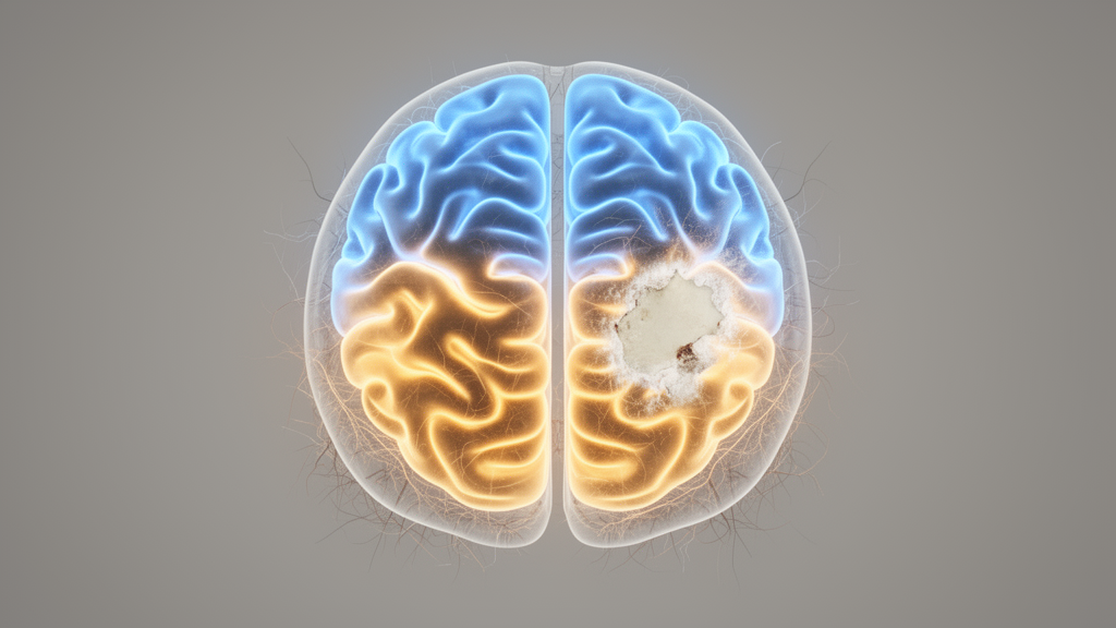
A unilateral ischemic stroke does not silence only the hemisphere it damages. In a rat study published in Behavioral and Brain Functions, researchers recorded neural activity from the caudal forelimb area on both sides of the brain and found widespread bilateral disruptions in oscillatory dynamics across delta, theta, alpha, beta, and gamma frequency bands after stroke.
The recordings combined several complementary analytical approaches. The researchers measured spectral power in canonical frequency bands, characterized the aperiodic 1/f slope of the local field potentials as an index of excitation–inhibition balance, and assessed cross-frequency coupling between the two hemispheres as a marker of interhemispheric coordination.
Two findings stand out. First, the aperiodic 1/f slope shifted after stroke, which the authors interpret as reflecting an altered balance between excitatory and inhibitory neural activity.
Second, cross-frequency coupling between hemispheres, particularly the coupling of low-frequency phase to gamma amplitude, was reorganized after the stroke. The team reports that this reorganization indicates impaired interhemispheric coordination and a bilateral imbalance in the motor network. In other words, the hemisphere that never experienced the infarct shows measurable changes in how its rhythms interact with those of the damaged side.
The authors conclude that post-stroke motor dysfunction arises from disrupted, state-dependent dynamics of distributed motor networks rather than from isolated changes confined to the lesioned hemisphere.
An important limitation deserves emphasis. The main experimental cohort did not include a sham-operated control group. However, a pilot experiment with three sham and three stroke animals, conducted using identical procedures, provided supportive evidence that the key measures remain stable under sham conditions. Readers should therefore treat the sham comparison as preliminary rather than definitive, and the anesthetized recordings add a further caveat about how the observed dynamics translate to awake behavior, even though task-based recordings were also collected.
Because the work was conducted in rats, the findings are preclinical and cannot be directly extended to human patients or to therapy. Still, the multiband, bilateral picture they paint offers a technically detailed map of how a focal lesion reverberates through a distributed motor circuit.
Subject of Research: Bilateral electrophysiological disruptions in the rat caudal forelimb area after ischemic stroke
Article Title: Bilateral cortical network disruptions in the rat caudal forelimb area after unilateral stroke
Article References: Cao, C., Chen, D., Gong, J., Gao, S., Huang, J., Jia, C., Liu, H., Li, Y., & Guo, Y. (2026). Bilateral cortical network disruptions in the rat caudal forelimb area after unilateral stroke. Behavioral and Brain Functions. https://doi.org/10.1186/s12993-026-00355-9
Image Credits: AI Generated
DOI: 10.1186/s12993-026-00355-9
Keywords: ischemic stroke, caudal forelimb area, local field potentials, oscillatory dynamics, excitation–inhibition balance, cross-frequency coupling, interhemispheric coordination, motor network, rat model, MCAO, single-pellet retrieval, aperiodic 1/f slope
News Source: Cassandra Pierce. (October 29, 2026). Stroke Disrupts Brain Rhythms on Both Sides in Rat Motor Cortex. Scienmag.