When a bone fracture is fixed with an intramedullary nail and infection takes hold months later, the bacteria rarely confine themselves to the visible break. Pathogens can travel along the metal implant and the endosteal surface of the bone, seeding the nail entry site, the entire medullary canal, and the tracts drilled for the proximal and distal locking screws. A retrospective cohort study now reports that systematically debriding this entire nail-related infection pathway, followed by staged reconstruction, achieved infection control and bone union in nearly all of a carefully selected group of 47 patients treated at a single tertiary referral centre between January 2017 and January 2025.
The study, published in the Journal of Bone and Joint Infection, enrolled consecutive adults aged 18 to 60 with late fracture-related infection after tibial or femoral nailing. Every participant had fracture nonunion and a documented interval of at least three months from the first confirmed diagnosis of infection to admission. Infection was defined using the international consensus framework, requiring confirmatory evidence such as a sinus tract communicating with bone or implant, purulent drainage, phenotypically indistinguishable microorganisms from at least two deep specimens, or a histopathological threshold of more than five polymorphonuclear neutrophils per high-power field across at least five fields. The cohort comprised 39 men and eight women with a mean age of 40.2 years; 21 infections involved the tibia and 26 the femur, and the mean documented infection interval before admission was 15.3 months.
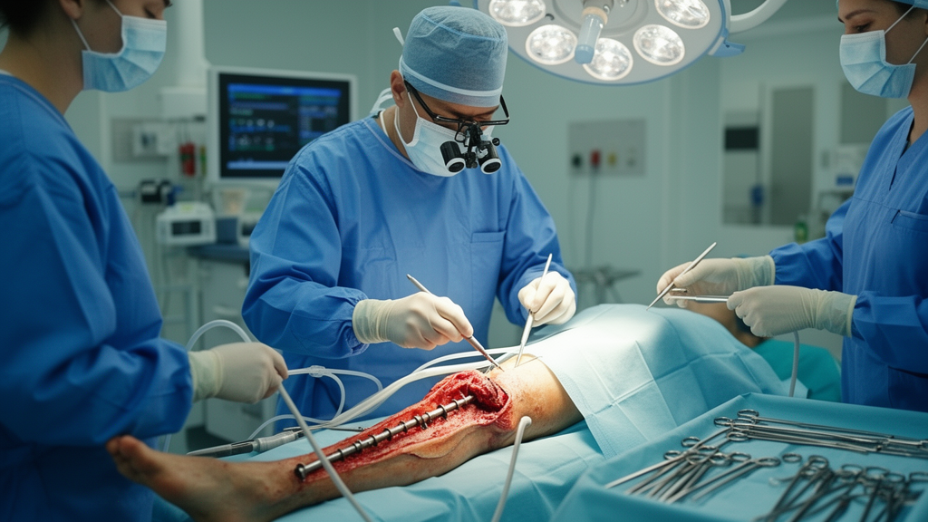
The treatment protocol unfolded in two deliberate stages. In Stage I, surgeons removed the intramedullary nail and performed systematic multi-regional debridement of the fracture and nonunion site, the nail entry site, the entire medullary canal, and all proximal and distal locking screw tracts. The canal was sequentially reamed to at least two millimetres beyond the removed nail’s diameter, and screw tracts were inspected along their full trajectories, enlarged and curetted when loosening, granulation tissue, or purulence was visible. In 14 patients, additional cortical windows or longitudinal grooves were created when the adequacy of intramedullary debridement could not be confirmed through the entry site and screw tracts alone. Irrigation and intermittent negative-pressure suction continued until the effluent ran clear of debris.
Stability was then restored with locking plate fixation, and dead space was managed with polymethyl methacrylate cement loaded with vancomycin at a fixed concentration of 5 grams per 40 grams of cement powder. The cement served multiple roles: a local Gram-positive antimicrobial carrier, a defect filler, and a spacer that permits an induced membrane to form around it, following the principles of the Masquelet technique. The intramedullary cement rod, moulded around a Kirschner wire core, was deliberately non-load-bearing; the locking plate carried the mechanical load. Soft-tissue defects were covered with flaps in four patients. Definitive reconstruction was not scheduled at a fixed interval but awaited clinical and laboratory quiescence of infection, no earlier than six to eight weeks after the first operation.
At Stage II, the temporary plate and all cement were removed, repeat debridement was performed, and the operative field was reassessed before bone grafting. Autologous iliac cancellous bone was grafted into the defect, supplemented with morselized allograft limited to no more than 25 percent when needed, and a new internal locking plate provided definitive fixation. No intramedullary nail was reinserted in any patient. The mean interval between stages was 11.4 weeks, with a range of 7 to 22 weeks, reflecting the individualized, reassessment-driven timing of the protocol.
The microbiological findings underscored how difficult these infections are to pin down. Deep-tissue cultures were positive in 30 patients and negative in 17, yet 15 of those culture-negative cases fulfilled clinical confirmatory criteria through communicating sinuses or intraoperative purulence, and the remaining two met the histopathological threshold. All 47 histopathology reports described changes compatible with osteomyelitis. Every patient received 42 days of systemic antimicrobial therapy after Stage I, beginning with empirical intravenous ceftriaxone and continuing with susceptibility-directed treatment; in culture-negative cases, the institutional regimen combined intravenous ceftriaxone with oral levofloxacin and rifampicin. Vancomycin-loaded cement was explicitly regarded as an adjunct rather than a stand-alone antimicrobial treatment, particularly because it does not reliably cover the Gram-negative organisms identified in the cohort.
The outcomes were striking, though tempered by important caveats. Two patients developed confirmed persistent infection requiring repeat debridement before definitive reconstruction, one diagnosed through repeated cultures yielding Staphylococcus aureus and the other through histopathology after suspicious intraoperative findings prompted deferral of bone grafting. Both completed a new 42-day antimicrobial course and proceeded to reconstruction. After definitive Stage II surgery, no confirmed recurrence and no unplanned debridement for suspected recurrence occurred during a mean follow-up of 25.3 months, ranging from 12 to 52 months. Union after the index reconstruction was achieved in 44 of 47 patients, or 93.6 percent; the remaining three achieved union after repeat bone grafting, yielding a final union rate of 100 percent.
Functional and laboratory measures moved in parallel. The mean time to radiographic union was 9.7 months and to unrestricted full weight-bearing 11.3 months. Median C-reactive protein fell from 26.5 to 6.3 milligrams per litre, erythrocyte sedimentation rate from 39.0 to 17.0 millimetres per hour, and white blood cell count from 7.2 to 5.9 times ten to the ninth per litre. Lower Extremity Functional Scale scores rose from a median of 47.0 to 70.0, Short Form-36 totals from 417.0 to 491.0, while pain and anxiety scores fell, all comparisons reaching statistical significance. Complications included delayed superficial donor-site wound healing in five patients, knee stiffness in five femoral cases, and limb length discrepancies of 2.7 to 4.3 centimetres in three patients.
The authors are careful about what these results do and do not demonstrate. This was a single-centre retrospective cohort without a comparator group, leaving it open to selection bias, residual confounding, and indication bias, and it cannot establish superiority over alternative strategies such as nail retention or antimicrobial suppression. Microbiological procedures in Stage I were not fully standardized: four patients had only two deep specimens, separate instruments were not routinely used, aerobic incubation lasted just five days, and anaerobic culture was selective, factors that may have contributed to the culture-negative proportion. Sampling at the planned Stage II procedure was selective rather than systematic, so subclinical residual infection cannot be excluded, and the absence of clinical recurrence should not be read as culture-confirmed eradication. The follow-up period may also have been too short to capture all late recurrences.
Within those limits, the study offers a concrete technical template for one of orthopaedic infection’s most stubborn presentations: late infection with nonunion after intramedullary nailing, where mature biofilm and necrotic bone make implant retention unlikely to succeed. The distinguishing feature was not the Masquelet-style reconstruction itself but the systematic extension of debridement beyond the fracture site to the entire implant pathway, a rationale grounded in the known propensity of pathogens to spread along nails and endosteal surfaces. The authors conclude that the protocol is feasible in selected patients and call for multicentre comparative studies to test whether whole-pathway debridement genuinely outperforms more limited approaches.
Subject of Research: Staged multi-regional debridement and reconstruction for late fracture-related infection after intramedullary nailing
Article Title: Multi-regional debridement and staged reconstruction for fracture-related infection after intramedullary nailing
Article References: Chen, X., Li, J., Li, Y., Li, H., Lv, K., Zhu, Y., Song, M., & Shi, J. (2026). Multi-regional debridement and staged reconstruction for fracture-related infection after intramedullary nailing. Journal of Bone and Joint Infection, 11(5), 655-665. https://doi.org/10.5194/jbji-11-655-2026
Image Credits: AI Generated
Keywords: fracture-related infection, intramedullary nailing, osteomyelitis, nonunion, debridement, Masquelet technique, vancomycin-loaded cement, locking plate fixation, staged reconstruction, bone grafting, retrospective cohort, orthopaedic surgery
News Source: Kristina Jarvis. (October 29, 2026). Staged Debridement Protocol Controls Late Infection After Intramedullary Nailing. Scienmag.