Recent advancements in pediatric radiology have shed light on the challenging landscape of vertebral tumors in children. These rare but significant conditions can often be misdiagnosed or overlooked due to their atypical presentations. A comprehensive study elucidates the radiologic features of these tumors, underscoring the necessity for accurate differentiation between various types of tumors and other spinal anomalies. Understanding these differences is crucial for effective treatment and management strategies tailored specifically for the pediatric population.
The complexity of diagnosing pediatric vertebral tumors lies in the fact that many present with nonspecific symptoms, which can mimic more benign conditions like infections or trauma. Symptoms such as back pain or neurological deficits may lead physicians down a convoluted diagnostic pathway. Thus, it’s paramount for medical professionals to have a keen understanding of typical imaging characteristics associated with various tumors. This study provides invaluable insights into the imaging techniques that can aid radiologists in making accurate diagnoses.
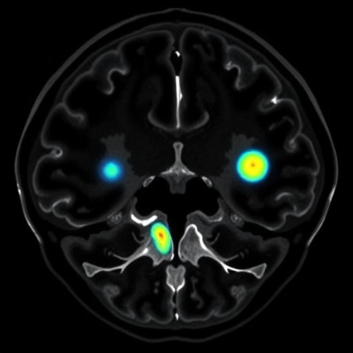
Radiologic imaging, including MRI and CT scans, plays a pivotal role in identifying vertebral tumors. MRI, in particular, is superior due to its ability to provide detailed images of soft tissue structures and spinal cord involvement. In the study, MRI findings revealed key characteristics of different tumor types, such as osteoblastoma and Ewing sarcoma. Each of these tumors has unique imaging signatures that, when recognized, can significantly expedite the diagnosis process and improve patient outcomes.
Moreover, the differential diagnoses between benign and malignant vertebral tumors are crucial for formulating a treatment plan. Benign tumors like hemangiomas may require less aggressive treatment compared to malignant types that may necessitate chemotherapy or surgical intervention. Understanding the radiological distinctions—such as the presence of a soft tissue mass or vertebral body destruction—can lead to more precise and timely interventions. This is particularly essential for pediatric patients, whose treatment pathways can differ significantly from adults.
The paper highlights specific radiologic features that correlate with histological findings, further emphasizing the role of imaging in guiding clinical decision-making. For example, the presence of a specific pattern of bone marrow edema on MRI can hint at inflammatory processes versus neoplastic growth. By recognizing these patterns early, healthcare providers can better tailor the clinical approach, potentially leading to improved morbidity and mortality rates in affected children.
Diagnostic imaging not only provides clarity about the tumor itself but also sheds light on its extent and the associated risks. Advanced imaging techniques can reveal important anatomical relationships, such as involvement of surrounding structures, which is crucial in surgical planning. Protecting the spinal cord and surrounding nerves during intervention is critical, given the potential for debilitating outcomes if incorrect approaches are taken.
Understanding the age-related variations in presentations of vertebral tumors in pediatric populations is an additional layer of complexity. Tumor behavior can vary greatly depending on the age at diagnosis, as younger patients may exhibit different biological behaviors compared to adolescents. These nuances should be carefully considered by radiologists and clinicians in their assessments and during the treatment planning process.
In addition, the study places significance on collaborative conversations among multidisciplinary teams that include radiologists, pediatric oncologists, and spine surgeons. This collaboration fosters a more comprehensive approach to managing vertebral tumors, ensuring that all aspects of a child’s health are being considered. Shared decision-making also enhances the patient experience, making it essential for healthcare providers to engage families in the treatment planning process.
Emerging technologies, such as artificial intelligence, are being integrated into radiology practices to enhance the diagnostic process further. AI-driven models can assist in recognizing patterns in imaging studies that may be subtle and easily missed by the human eye. These technologies are not meant to replace human judgment but serve as tools to augment capabilities, allowing radiologists to focus on more complex cases that require careful interpretation.
As this field continues to evolve, educational efforts aim to bridge gaps in knowledge among healthcare professionals regarding pediatric vertebral tumors. Continued dissemination of research findings through journals and conferences is vital to keep practitioners informed about the latest radiologic features and treatment protocols. This will ultimately lead to better outcomes for young patients grappling with these challenging diagnoses.
Finally, ongoing research into biological markers and their relationship with imaging characteristics is imperative. Such investigations could unveil new frontiers in personalized medicine. By correlating radiologic features with molecular and genetic markers, there may soon be the potential to predict tumor behavior, effectively guiding treatment strategies even before histological evaluations are performed.
In culmination, the intricate nature of pediatric vertebral tumors underscores the evolving landscape of pediatric radiology. The importance of precise imaging techniques cannot be overstated, as early and accurate diagnosis is essential for optimizing treatment outcomes. As research in this field progresses, the integration of innovative technologies and interdisciplinary cooperation will pave the way for even greater advancements in the management of these complex and often devastating diseases in children.
Subject of Research: Pediatric Vertebral Tumors
Article Title: Pediatric vertebral tumors: radiologic features and differential diagnoses.
Article References:
Inarejos Clemente, E., Navallas, M., Ladera, E. et al. Pediatric vertebral tumors: radiologic features and differential diagnoses.
Pediatr Radiol (2025). https://doi.org/10.1007/s00247-025-06488-9
Image Credits: AI Generated
DOI: 15 December 2025
Keywords: Pediatric vertebral tumors, radiologic features, differential diagnoses, MRI, CT scans, pediatric oncology, bone tumors, multidisciplinary care, artificial intelligence in radiology, personalized medicine.
Tags: advancements in pediatric tumor imagingchallenges in diagnosing pediatric tumorsdifferential diagnosis of spinal tumorsimaging techniques in pediatric radiologyimportance of accurate tumor identificationmanagement strategies for pediatric tumorsmisdiagnosis of vertebral tumorsMRI and CT scans for tumorspediatric vertebral tumorsradiologic features of spinal anomaliesspinal cord involvement in tumorssymptoms of vertebral tumors in children


