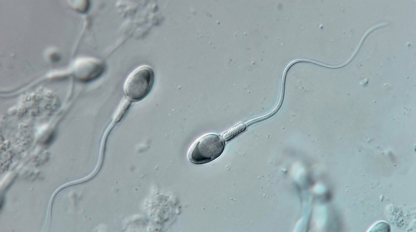
Men with high levels of sperm DNA fragmentation who underwent assisted reproduction at a single Chinese fertility center had markedly poorer semen quality and a lower rate of normal fertilization than men with lower fragmentation, but their chances of achieving pregnancy and live birth after embryo transfer were not significantly different, according to a retrospective cohort study published in Basic and Clinical Andrology. The findings, drawn from 1,784 IVF and ICSI cycles performed between January 2020 and January 2022, add real-world evidence to a long-running debate over how much weight the sperm DNA fragmentation index deserves in the clinical workup of infertile couples.
The sperm DNA fragmentation index, or DFI, measures the proportion of sperm carrying breaks in their DNA strands. It has emerged in recent years as a potential marker of male fertility that goes beyond the traditional semen analysis of volume, concentration, motility and morphology. The sixth edition of the World Health Organization laboratory manual for semen examination describes DFI testing as an important supplement to standard semen analysis, yet conflicting results across studies have led some guidelines to refrain from recommending routine DFI testing in infertility evaluations. The authors of the new study, led by Chaofeng Wei and Qihui Liang with colleagues at the Affiliated Hospital of Shandong University of Traditional Chinese Medicine, set out to quantify the association between DFI and conventional semen parameters, embryological outcomes and pregnancy outcomes while rigorously adjusting for confounders.
The team analyzed data from patients who underwent IVF or ICSI with embryo transfer at the hospital’s Reproduction and Genetics Center, drawing on records in the Reproductive Case Management System. Eligible cycles were those in which the male partner had undergone DFI testing by sperm chromatin structure analysis, a flow-cytometry method in which sperm DNA is partially denatured with an acid-detergent solution and stained with acridine orange; the ratio of red, denatured single-stranded DNA fluorescence to total fluorescence yields the fragmentation index. At least 5,000 sperm cells were captured per sample, with measurements repeated to ensure reproducibility. The researchers excluded couples with chromosomal abnormalities, polycystic ovary syndrome, endometriosis, recurrent miscarriage or repeated implantation failure, uterine malformations, endocrine diseases such as diabetes and thyroid disease, and mixed or rescue-ICSI cycles, in order to limit confounding from female factors.
Of the 1,784 first IVF or ICSI cycles included, 1,652 had a normal DFI below 30 percent and 132 had a high DFI at or above that threshold. Men in the high-DFI group were older on average, at 36.89 versus 34.58 years, had smaller testicular volumes and a higher prevalence of varicocele. Their semen quality was significantly impaired: sperm concentration averaged 43.55 versus 77.40 million per milliliter, progressive motility was 25.65 versus 39.20 percent, and total motility was 35.80 versus 55.49 percent, all differences statistically significant at P < 0.001. Sperm morphology and semen volume, by contrast, did not differ significantly between the groups.
Simple linear regression revealed moderate negative correlations between DFI and progressive motility and between DFI and total motility, a slight negative correlation with sperm concentration, and no significant correlation with semen volume or morphology. After adjusting for male age, varicocele status and testicular volume in multivariate linear regression, high DFI remained independently associated with lower sperm concentration, with a coefficient of −31.15 (95 percent confidence interval −44.75 to −17.55), lower progressive motility (B = −12.21, 95 percent CI −14.87 to −9.56) and lower total motility (B = −18.00, 95 percent CI −21.38 to −14.62), all at P < 0.001. Morphology, analyzed with multiple imputation to handle roughly 29 percent missing data, showed no significant association with DFI. The authors caution that the retrospective design limits causal inference: poor motility could plausibly affect DFI rather than result from it, or both could reflect a shared underlying problem such as elevated reactive oxygen species or mitochondrial damage.
The study then turned to laboratory outcomes of the ART cycles. Unadjusted 2PN fertilization rates, calculated as the number of normally fertilized oocytes with two pronuclei divided by the total number of retrieved oocytes, showed no significant difference between groups, with medians of 0.636 in the normal-DFI group and 0.592 in the high-DFI group. However, after adjusting for female age, male age, estradiol level on the trigger day and insemination mode in a beta regression model suited to bounded proportions, high DFI was significantly associated with a reduced 2PN fertilization rate, with a coefficient of −0.257 (95 percent CI −0.491 to −0.024, P = 0.031). No significant differences emerged in the proportion of patients with at least one top-quality day-3 embryo or with more than 50 percent transferable embryos, either unadjusted or after logistic regression adjustment.
Pregnancy outcomes were assessed in a subset of 736 fresh embryo transfer cycles, after excluding cycles without transferable embryos or involving full embryo cryopreservation, to avoid confounding from freezing and thawed-transfer strategies. In this subset the high-DFI group again had older female and male partners, lower motility and a higher proportion of ICSI cycles. Yet unadjusted and adjusted comparisons showed no significant differences between groups in biochemical pregnancy, clinical pregnancy, live birth, pregnancy loss or ectopic pregnancy, all with P values above 0.05. The authors suggest that sperm preparation procedures such as density gradient centrifugation and the swim-up method, which remove large numbers of poorly motile sperm before insemination, may have diminished any impact of high DFI on pregnancy outcomes, and they note that paternal DNA damage may be partially repaired by the oocyte after fertilization, since all DNA repair in pre-implantation embryos relies on maternal contribution.
Sensitivity analyses largely supported the main results. A zero-one inflated beta regression model, designed to handle the clustering of proportion data at the boundaries of 0 and 1, placed the posterior distribution of the DFI effect on the 2PN rate in the negative region, with a posterior mean of −0.103 and an 89.1 percent probability that high DFI reduces the fertilization rate, though the 95 percent credible interval of −0.267 to 0.063 crossed zero. Propensity score matching for pregnancy outcomes and alternative DFI cut-offs of 25 and 35 percent yielded results consistent with the primary findings, with the reduced 2PN fertilization rate persisting at both alternative thresholds. Restricted cubic spline analysis of DFI as a continuous variable found a marginally significant linear downward trend for the 2PN fertilization rate and a statistically significant non-linear relationship for live birth, with the curve dropping more steeply once DFI exceeded the 30 percent range, a pattern the authors interpret as a possible threshold effect that binary comparisons may miss when the high-risk group is diluted by borderline cases.
The authors acknowledge several limitations. The relatively small number of ICSI cycles precluded subgroup analyses by insemination method, although insemination mode was included as a covariate. Excluding patients with recurrent miscarriage or repeated implantation failure strengthened internal validity but may limit external validity by omitting a high-risk subpopulation in which DFI could exert a stronger effect. The high-DFI group in the fresh transfer cohort was small, which constrained statistical power for low-frequency events such as miscarriage and raised the risk of overfitting in the regression models, so findings on clinical outcomes should be interpreted with caution. Blastocyst formation was not assessed because nearly half of the cycles did not undergo extended embryo culture, and restricting analysis to selected cycles could have introduced selection bias.
Overall, the study concludes that high sperm DFI is independently associated with poorer semen quality and a reduced 2PN fertilization rate but was not significantly associated with embryo quality or pregnancy outcomes in this cohort once female and cycle-related factors were taken into account. The authors say the findings support the clinical relevance of DFI in evaluating male fertility potential while suggesting its limited predictive value for ART success, and they call for large-scale prospective studies integrating molecular mechanisms with rigorous statistical modeling to better define the role of sperm DNA fragmentation in assisted reproduction.
Subject of Research: Association between sperm DNA fragmentation index and semen quality, embryological outcomes and pregnancy outcomes in assisted reproductive technology
Article Title: Association of sperm DNA fragmentation index with semen quality and ART outcomes: real-world evidence from a retrospective cohort
Article References: Wei, C., Liang, Q., Liu, D., Yu, Y., Wei, Y., Li, Y., Wu, H., & Lian, F. (2026). Association of sperm DNA fragmentation index with semen quality and ART outcomes: real-world evidence from a retrospective cohort. Basic and Clinical Andrology, 36(1), Article 9. https://doi.org/10.1186/s12610-026-00308-2
Image Credits: AI Generated
DOI: 10.1186/s12610-026-00308-2
Keywords: sperm DNA fragmentation, DFI, male infertility, semen quality, IVF, ICSI, 2PN fertilization rate, embryo quality, live birth, retrospective cohort, assisted reproductive technology, sperm chromatin structure analysis
News Source: Ophelia Keating. (November 5, 2026). Sperm DNA Damage Linked to Poorer Semen Quality and Lower Fertilization Rate in IVF Study. Scienmag.