A heart transplant can restore circulation, reverse advanced heart failure and give patients years of life, but the new organ remains under constant surveillance by the immune system. The recipient’s body recognizes the transplanted heart as biologically foreign, creating the possibility of cardiac allograft rejection. Clinicians currently rely on tissue biopsies, imaging, blood tests and clinical symptoms to detect this process, yet rejection is not a single event driven by one cell type. It is a changing biological program involving immune cells, blood vessels, stromal tissue and the heart muscle itself. A study published in Nature Cardiovascular Research now uses spatial transcriptomics to examine that process directly inside human cardiac transplant tissue, revealing how rejection-related cellular activity is organized across the architecture of the graft.
The work by Amancherla, Oill, Bledsoe and colleagues addresses a central limitation in transplant biology: conventional molecular analyses often remove tissue from its spatial context. When a biopsy is homogenized, RNA from thousands of cells is mixed together, allowing researchers to measure which genes are active but not precisely where that activity occurs. Spatial transcriptomics preserves the physical coordinates of gene expression. In practical terms, it can show whether a particular immune program is concentrated around a blood vessel, embedded within the myocardium, positioned at the boundary between scar tissue and viable muscle, or distributed across the graft. That information is essential because neighboring cells can influence one another through direct contact, secreted cytokines and changes to the local extracellular matrix.
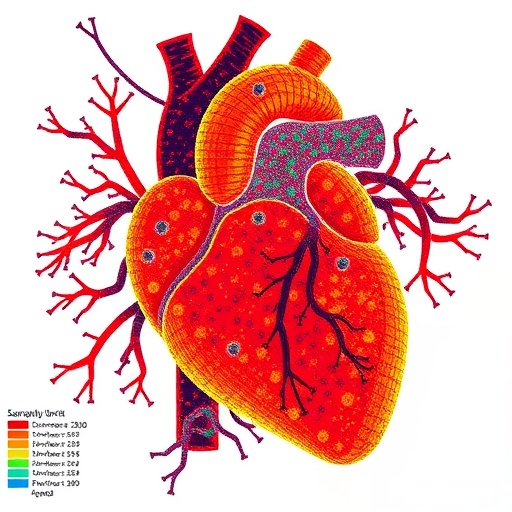
At the technical level, spatial transcriptomics links molecular measurements to tissue morphology. Thin sections of the transplanted heart are placed on arrays containing thousands of spatially barcoded capture regions. Messenger RNA released from the tissue is collected together with the positional barcode of the region from which it originated. Sequencing then produces a map in which each coordinate contains a partial gene-expression profile. Computational analysis can classify cellular states, identify genes associated with inflammation or tissue injury, and reconstruct neighborhoods in which several cell populations operate together. Unlike a conventional microscope image, the resulting map can reveal molecular changes before they become obvious as structural damage. Unlike single-cell RNA sequencing alone, it retains the geography needed to understand local interactions.
The study’s significance lies in its treatment of rejection as a dynamic process rather than a static label. A biopsy reported as “rejection” may contain several overlapping phases: immune recognition, recruitment of circulating leukocytes, activation of vascular cells, injury to cardiomyocytes and an attempt by the tissue to repair itself. These phases can coexist in different regions of the same graft. Spatially resolved analysis makes it possible to distinguish a focal inflammatory microenvironment from a more diffuse response and to identify whether the most active programs are located in the vascular compartment, the interstitium or the contractile tissue. The authors’ approach therefore moves beyond simply asking which genes are elevated and instead asks where, when and in which cellular neighborhood those genes become active.
One important biological theme in cardiac allograft rejection is communication between immune cells and the graft’s vascular system. Endothelial cells line the blood vessels and form the first major interface between circulating immune cells and transplanted tissue. Under inflammatory conditions, they can alter adhesion molecules, chemokines and antigen-presentation pathways, making it easier for immune cells to exit the bloodstream and enter the heart. Spatial transcriptomics can detect these endothelial changes alongside the immune populations gathered around them. This provides a mechanistic view of how a localized vascular signal may become a broader tissue response. It also helps explain why microvascular injury can be an early and clinically important feature of rejection, even when damage to the heart muscle is not yet extensive.
The mapped cellular programs also illuminate the roles of immune populations that are often grouped together in routine pathology. T lymphocytes, macrophages, antigen-presenting cells and other leukocytes do not perform identical functions, and their effects depend on their activation state and location. Some may recognize donor-derived antigens and coordinate direct attack, while others release inflammatory mediators, clear damaged cells or promote tissue remodeling. The surrounding fibroblasts and endothelial cells are not passive observers: they can respond to immune signals, change the composition of the extracellular matrix and influence whether inflammation resolves or becomes persistent. By measuring gene-expression patterns across these interacting populations, the study frames rejection as a multicellular circuit rather than an isolated assault by immune cells.
This distinction could be particularly valuable for understanding why transplant recipients experience variable clinical courses. Two biopsies may appear broadly similar under the microscope while representing different molecular trajectories. One may reflect an active immune response that could be rapidly controlled with immunosuppressive treatment; another may show residual inflammation, vascular stress or repair-associated remodeling after the peak of rejection has passed. Spatial gene-expression signatures could eventually help separate these states. They may also reveal molecular activity in tissue that looks only mildly abnormal by conventional histology, offering a potential route toward earlier detection. The study does not by itself replace established clinical diagnostics, but it supplies a high-resolution framework for testing whether specific spatial programs correlate with outcomes, treatment response or later graft dysfunction.
The findings are also relevant to the long-term problem of chronic cardiac allograft injury. Acute rejection is only one threat to a transplanted heart. Over time, repeated immune activation and vascular damage can contribute to cardiac allograft vasculopathy, a progressive narrowing of the coronary vessels that limits blood flow and can lead to graft failure. Chronic injury involves interactions among endothelial cells, immune populations, smooth-muscle cells and fibroblasts, along with structural changes in the vessel wall. A spatial approach may help connect early inflammatory states with later remodeling. If researchers can identify cellular arrangements that consistently precede vascular disease, those signatures could become targets for monitoring or intervention before irreversible anatomical changes develop.
The research also demonstrates why human tissue is indispensable in transplant science. Animal models have provided foundational knowledge about alloimmunity, but human grafts exist within distinct clinical histories, immunosuppressive regimens, infections and episodes of prior injury. Those factors can reshape cellular states in ways that are difficult to reproduce experimentally. Spatial transcriptomics allows investigators to study this complexity while maintaining the anatomical context of the patient-derived specimen. The approach can be combined with histology, immunostaining, single-cell sequencing and clinical records, creating a layered view of rejection that links molecules to cells, cells to tissue regions and tissue patterns to patient outcomes. As larger datasets become available, computational models may be able to classify rejection states with increasing precision.
The broader message from the study is that transplanted organs are not biologically uniform landscapes. Within a small piece of myocardium, immune activation, vascular signaling, tissue injury and repair may occupy distinct but connected territories. Mapping those territories offers a more realistic picture of how rejection develops and why its consequences vary across patients. The work points toward a future in which transplant surveillance is guided not only by the presence or absence of inflammation, but also by the identity, location and trajectory of the programs driving it. For heart-transplant medicine, that could ultimately support more individualized immunosuppression—strong enough to protect the graft, yet restrained enough to reduce infection, malignancy and drug toxicity. By revealing the cellular geography of human cardiac allograft rejection, the study turns a clinically familiar diagnosis into a dynamic molecular story.
Subject of Research: Human cardiac allograft rejection and the spatially organized cellular programs involved in transplanted-heart injury.
Article Title: Dynamic cellular programs of human cardiac allograft rejection revealed by spatial transcriptomics
Article References: Amancherla, K., Oill, A.M.T., Bledsoe, X. et al. Dynamic cellular programs of human cardiac allograft rejection revealed by spatial transcriptomics. Nature Cardiovascular Research (2026). https://doi.org/10.1038/s44161-026-00849-9
Image Credits: AI Generated
DOI: https://doi.org/10.1038/s44161-026-00849-9
Keywords: cardiac transplantation, cardiac allograft rejection, spatial transcriptomics, human heart, transplant immunology, immune-cell interactions, endothelial cells, cardiac allograft vasculopathy, tissue microenvironments, precision medicine
Tags: advancements in spatial transcriptomics for transplant researchblood vessel and stromal tissue involvement in rejectioncellular programs in cardiac allograft rejectiongene expression mapping in cardiac tissueimmune cell infiltration patterns in heart graftsimmune cell localization in transplanted heartsimmune response spatial organization in heart transplantslimitations of traditional biopsy methodsmolecular analysis of transplant rejectionspatial transcriptomics in heart transplant rejectiontissue architecture in transplant rejectionunderstanding rejection mechanisms through spatial gene expression