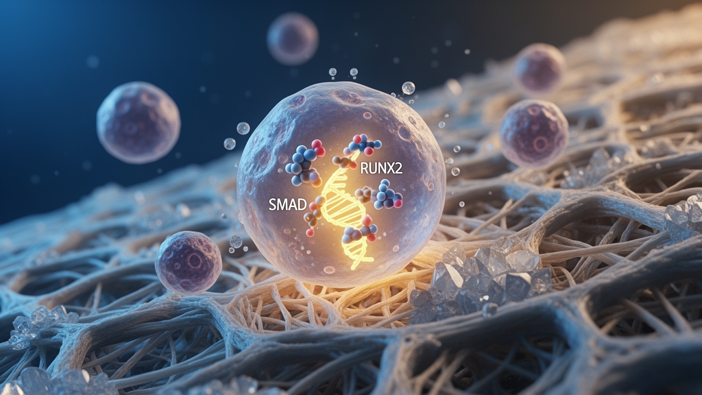
Researchers have uncovered how dynamic molecular complexes between osteogenic transcription factor RUNX2 and receptor-regulated SMAD proteins control breast cancer bone metastasis. Bone is the most common site of breast cancer spread, causing clinical complications including pain, pathological fractures, hypercalcemia, spinal cord compression, and impaired mobility, while driving secondary dissemination to other organs. Historically, Runt-related transcription factor 2 (RUNX2) was viewed as a downstream target of SMAD proteins during osteoblast differentiation and bone metastasis. The newly published study demonstrates that RUNX2 does not merely accept upstream instructions; instead, physical SMAD–RUNX2 complexes actively amplify signaling throughput of transforming growth factor-beta and bone morphogenetic protein pathways. Utilizing cancer models, molecular mutagenesis, transcriptomic profiling, and rodent assays, the team showed that TGFβ-induced SMAD2/3–RUNX2 complexes and BMP-activated SMAD1–RUNX2 complexes carry out distinct, microenvironment-dependent tasks across metastatic progression. Mechanistically, competitive binding between SMAD2/3 and SMAD1 for the same structural domain on RUNX2 governs crosstalk between these two cascades.
Structurally, SMAD proteins interact with the SMAD-interacting domain (SMID) of RUNX2, located within the nuclear matrix-targeting signal domain spanning amino acids 391 to 432. In normal skeletal physiology, SMAD1, SMAD5, and SMAD8 transduce BMP signals to stimulate osteoblast differentiation and chondrocyte maturation, whereas SMAD2 and SMAD3 transduce TGFβ signals to attenuate osteogenesis by downregulating osteogenic genes. To examine how these complexes operate in malignancy, investigators utilized human breast cancer cell lines MDA-MB-231, BT549, and MCF7, alongside osteoblast-like MG-63 cells. Cell lines were authenticated by short tandem repeat profiling and routinely tested for mycoplasma contamination. Researchers engineered lentiviral vectors encoding Flag-tagged fusions of full-length wild-type RUNX2, an R391A point-mutant termed RUNX2 MU, and a truncated variant designated RUNX2 ∆C lacking the SMID domain from residue 391 onward. Following transduction and puromycin selection, exogenous RUNX2 expression triggered feedback inhibition of endogenous RUNX2 transcripts, allowing the authors to directly assess how structural alterations modulate cellular responses to cytokine exposure.
Biochemical characterization through coimmunoprecipitation confirmed that recombinant TGFβ1 and TGFβ3 stimulated phosphorylation and nuclear translocation of SMAD2/3, forming stable complexes with Flag-tagged wild-type RUNX2 in MDA-MB-231 and BT549 cells. In parallel, recombinant BMP2 and BMP7 stimulated phosphorylation and nuclear accumulation of SMAD1, driving assembly with wild-type RUNX2. These interactions were impaired in cancer cells expressing the R391A mutant or SMID truncation variant. To determine functional consequences on gene transcription, the team conducted dual-luciferase reporter assays using a pGL3 plasmid containing a 6× osteoblast-specific cis-acting element, a 6×SMAD2/3-responsive reporter, and RT-qPCR quantification of the SMAD1 target gene COL1A1. While wild-type RUNX2 overexpression alone caused only modest baseline gains in reporter readouts, assembly of either SMAD2/3–RUNX2 or SMAD1–RUNX2 complexes significantly amplified RUNX2-mediated transcriptional output. Concurrently, these complexes amplified SMAD2/3 and SMAD1 transcriptional activity upon respective cytokine stimulation, confirming that physical complex formation functions as a mutual, bidirectional transcriptional booster in breast cancer cells.
To profile genome-wide transcriptional alterations driven by these complexes, investigators performed RNA sequencing on BT549 cells overexpressing wild-type RUNX2 or SMID variants following 48 hours of treatment with TGFβ1, TGFβ3, BMP2, BMP7, or phosphate-buffered saline. Principal component analysis revealed that cytokine treatments triggered widespread transcriptomic remodeling exclusively in wild-type RUNX2 cells, while exerting muted shifts in cells expressing RUNX2 MU or RUNX2 ∆C variants. Calculating standardized z-scores demonstrated significant upregulation of Reactome RUNX2-regulated pathways, Gene Ontology TGFβ-responsive signatures, and BMP-responsive signatures upon ligand exposure only when RUNX2 possessed an intact SMID domain. Differential expression analysis identified 254 unique genes responsive to TGFβ stimulation and 251 unique genes responsive to BMP stimulation specifically in wild-type cells. Functional enrichment showed that the TGFβ-induced SMAD2/3–RUNX2 complex activated genes governing motility, mesenchymal differentiation, and hypoxic adaptation, whereas the BMP-activated SMAD1–RUNX2 complex stimulated programs associated with epithelial differentiation, focal adhesion, and bone development while repressing mesenchymal and migratory genes.
Western blotting confirmed that transcriptional reprogramming translated into divergent protein expression patterns governing dissemination and niche colonization. Cells stimulated with TGFβ ligands exhibited substantial increases in integrin alpha 5 (ITGA5), an adhesion molecule facilitating breast cancer cell homing to bone matrix components, whereas BMP exposure caused no alteration in ITGA5 protein levels. Simultaneously, both complexes upregulated osteopontin (OPN) and bone sialoprotein (BSP), which represent canonical RUNX2 targets, as well as osteoblast adhesion molecule cadherin 11 (CDH11) and osteoclast activator RANKL, alongside genes promoting proliferation and suppressing apoptosis. In vitro Transwell assays demonstrated that wild-type RUNX2 enhanced cancer cell migration toward osteoblast-like MG-63 cells across an 11.5-hour window. Treatment with TGFβ1 and TGFβ3 significantly increased this osteotropic migration, whereas exposure to BMP2 and BMP7 inhibited migration. Furthermore, shRNA-mediated co-knockdown of SMAD2 and SMAD3 in wild-type cells weakened the promigratory effect of TGFβ ligands, confirming that SMAD2/3 transducers are functionally indispensable for driving RUNX2-dependent breast cancer osteotropism.
The study established that competitive binding of SMAD2/3 and SMAD1 to the RUNX2 SMID domain provides the molecular basis for antagonistic crosstalk between TGFβ and BMP cascades. In coimmunoprecipitation assays where MDA-MB-231 and BT549 cells received constant TGFβ1/3 concentrations alongside escalating BMP2/7 doses, rising BMP levels progressively displaced phosphorylated SMAD2/3 from RUNX2, blunting TGFβ-driven cancer cell migration toward osteoblasts. Reciprocally, holding BMP2/7 levels constant while escalating TGFβ1/3 concentrations dose-dependently reduced SMAD1–RUNX2 assembly and suppressed BMP-induced cellular behaviors. This competitive equilibrium also governed cancer cell adhesion to osteoblasts. In assays where GFP-labeled cancer cells were incubated over confluent MG-63 osteoblasts for 15 to 25 minutes, BMP treatment significantly increased adhesion efficiency. Disruption of the SMID domain or shRNA knockdown of SMAD1 abrogated this BMP-stimulated adhesion gain, whereas TGFβ slightly decreased adhesion. Crucially, applying a gradient of TGFβ progressively dismantled BMP-mediated adhesion in a dose-dependent manner, illustrating how relative ligand concentrations shift cancer cells between adhesive attachment and migratory detachment.
To evaluate how these complexes influence tumor survival within the bone microenvironment, authors conducted anchorage-dependent colony formation assays using soft agar prepared with MG-63 conditioned medium supplemented with 10 percent fetal bovine serum over 14 days. In MDA-MB-231 and BT549 cultures, the BMP-activated SMAD1–RUNX2 complex drove the formation of substantially enlarged colony diameters, and in luminal MCF7 cells overexpressing wild-type RUNX2, BMP stimulation caused multiple individual colonies to aggregate into large, cohesive clusters. In sharp contrast, the TGFβ-activated SMAD2/3–RUNX2 complex stimulated the emergence of numerous small, dispersed colonies, markedly increasing total colony counts while reducing average diameter. Mutational disruption or deletion of the RUNX2 SMID domain, as well as targeted knockdown of SMAD1 or SMAD2/3, abolished these distinct cytokine-induced colony growth patterns. When cells were co-treated with both TGFβ and BMP ligands, competitive inhibitory effects emerged in soft agar, demonstrating that the SMAD1–RUNX2 complex supports large, consolidated osteolytic colonies, whereas the SMAD2/3–RUNX2 complex fosters scattered, diffuse micrometastatic survival within osteoblast-conditioned niches.
To measure recruitment to bone marrow, GFP-labeled MDA-MB-231 cells pretreated with cytokines for 48 hours were injected into the left cardiac ventricle of mice, followed by marrow harvest from femurs and tibias 24 hours later. Quantitative RT-qPCR analysis of GFP transcript levels demonstrated that TGFβ-pretreated cells achieved significantly higher bone marrow recruitment, whereas BMP pretreatment suppressed marrow homing. Furthermore, in four-week intratibial models sustained by weekly local injections of TGFβ1/3 or BMP2/7, X-ray imaging and hematoxylin and eosin staining demonstrated that both complexes significantly expanded osteolytic bone erosion areas compared to controls, verifying that both signaling axes actively drive osteolytic bone destruction once cancer cells colonize skeletal tissue.
Although both complexes accelerated osteolytic bone destruction, the TGFβ-induced SMAD2/3–RUNX2 complex uniquely drove extraosseous dissemination away from the primary skeletal site. Histological analysis of distant organs from intratibially injected mice revealed secondary metastatic lesions within lungs, liver, and lymph nodes specifically following TGFβ treatment, whereas BMP treatment confined metastatic growth strictly within the injected tibia. To validate these experimental findings in patient populations, authors interrogated clinical microarray datasets, including GSE2034, GSE2603, and bone metastatic dataset GSE14020, alongside Human Protein Atlas records. In primary breast cancer patients with high RUNX2 expression, tumors characterized by high TGFB and low BMP mRNA levels exhibited a significantly higher incidence of bone metastasis and reduced bone metastasis-free survival compared to tumors expressing balanced or low ligand levels. Similarly, patients whose primary tumors displayed high RUNX2 levels accompanied by a TGFβ-responsive and BMP-non-responsive transcriptomic signature showed elevated bone metastasis rates, whereas no survival divergence occurred among patients with low RUNX2 expression, confirming that high TGFβ signaling combined with suppressed BMP activity at the primary site facilitates skeletal dissemination.
The authors noted several experimental limitations, acknowledging that in vitro migration, adhesion, and soft agar assays simplify complex metastatic behaviors into isolated experimental parameters. Furthermore, animal assays captured discrete endpoints of recruitment, retention, osteolysis, and dissemination rather than continuously tracking real-time assembly of SMAD–RUNX2 complexes in living tissue over time. Despite these constraints, the investigation resolves long-standing questions regarding how transforming growth factor-beta and bone morphogenetic protein pathways exert opposing yet cooperative effects during breast cancer progression. By demonstrating that RUNX2 functions as an active bidirectional amplifier and molecular switch rather than a passive target, findings provide a mechanistic framework for developing context-dependent therapeutic interventions. At the primary tumor stage, targeted inhibition of the TGFβ cascade could suppress osteotropic migration and dissemination, whereas blocking BMP signaling might prove ineffective. Conversely, within established bone metastases, effective clinical management may require dual inhibition of both TGFβ and BMP pathways to simultaneously curb osteolytic degradation and prevent secondary organ dissemination, or the development of small molecules that disrupt the physical SMAD–RUNX2 interface.
Subject of Research: Molecular mechanisms of breast cancer bone metastasis mediated by SMAD–RUNX2 signaling complexes
Article Title: Dynamic SMAD–RUNX2 complexes orchestrate breast cancer bone metastasis
Article References: Meng, R., Fang, J.-N., Zhou, J., & Li, X.-Q. (2026). Dynamic SMAD–RUNX2 complexes orchestrate breast cancer bone metastasis. Experimental & Molecular Medicine. https://doi.org/10.1038/s12276-026-01883-0
Image Credits: AI Generated
DOI: 10.1038/s12276-026-01883-0
Keywords: breast cancer, bone metastasis, RUNX2, SMAD proteins, TGF-beta signaling, BMP signaling, osteolysis, cancer dissemination, Dynamic, SMAD, complexes, orchestrate
News Source: Nathaniel Bowman. (November 9, 2026). SMAD and RUNX2 Complexes Dynamically Control Breast Cancer Bone Metastasis. Scienmag.