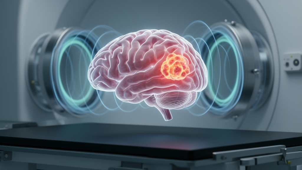
For decades, the gold standard for spotting brain tumors on magnetic resonance imaging has relied on a simple but consequential step: injecting a gadolinium-based contrast agent into the patient’s vein before the scan begins. Gadolinium brightens tumor tissue on T1-weighted images, sharpening the margins of lesions and making subtle masses easier to see. But the heavy metal has also accumulated a growing list of concerns, from the risk of nephrogenic systemic fibrosis in patients with kidney impairment to the well-documented deposition of gadolinium in brain tissue after repeated exposures. Now, a large retrospective study from Denmark suggests that for the critical task of screening, the contrast agent may not be necessary at all.
The research, conducted at Aalborg University Hospital in collaboration with Aarhus University Hospital and published in Frontiers in Neuroimaging, compared gadolinium-based contrast agent-free (GBCA-free) MRI against the full contrast-enhanced protocol in a real-world diagnostic pathway. The team included 191 patients harboring 195 intracranial tumors—124 intra-axial, arising within brain tissue itself, and 71 extra-axial, growing outside the brain parenchyma—alongside 410 randomly selected controls drawn from nearly 2,000 patients referred with suspected brain tumors between 2013 and 2022. The design mirrored the clinical reality of the North Denmark Region, where patients meeting national criteria for possible brain tumors are referred directly for MRI, typically within six days.
Three senior neuroradiologists independently reviewed anonymized scans, blinded to all clinical and demographic information. Each scan was first evaluated using only GBCA-free sequences: axial diffusion-weighted imaging with b-values of 0 and 1,000, sagittal T2-weighted imaging, and coronal T2-FLAIR. These sequences exploit the biophysical signatures of tumors—restricted water diffusion in densely cellular masses, prolonged T2 relaxation in edematous tissue, and abnormal signal on FLAIR that suppresses cerebrospinal fluid to highlight lesions near fluid-filled spaces. Only afterward were the same readers shown the complete dataset, now including T1-FLAIR images acquired seven and ten minutes after injection of 0.2 milliliters per kilogram of gadoteridol, in a randomized order designed to prevent recall bias.
The results upended expectations. GBCA-free MRI achieved a sensitivity of 79.6 percent and a specificity of 96.1 percent across the combined evaluation of all three raters. The contrast-enhanced protocol, traditionally considered the gold standard, performed slightly worse: a sensitivity of 73.3 percent and a specificity of 92.9 percent. The difference was not statistically significant, with a p-value of 0.15 and an odds ratio of 1.706 whose 95 percent confidence interval spanned 1. Agreement between the two approaches was substantial, measured by Cohen’s kappa at 0.699—a level of concordance that indicates the two methods reliably identify the same patients as tumor-positive or tumor-negative.
Subgroup analyses revealed comparable performance for intra-axial tumors, where GBCA-free imaging reached a sensitivity of 79.0 percent and specificity of 98.5 percent, outpacing the contrast-enhanced protocol’s 74.2 percent and 96.2 percent. For extra-axial tumors, both modalities showed modest sensitivity—56.3 percent for GBCA-free and 57.8 percent for contrast-enhanced—despite high specificity above 95 percent. The authors attribute these lower rates partly to the absence of clinical context during the blinded review: without knowing the patient’s symptoms or having access to prior imaging, radiologists were less inclined to label ambiguous hyperintensities as neoplastic. In routine practice, interpretation is embedded in a multidisciplinary discussion that typically raises detection rates.
The slightly superior sensitivity of the non-contrast approach may seem counterintuitive, but the authors propose a known perceptual pitfall as a possible explanation. When radiologists begin by examining contrast-enhanced sequences and find no abnormal enhancement, their detection efficiency for the non-contrast images that follow can decline—a phenomenon related to the satisfaction of search error, which occurs in roughly 22 percent of diagnostic radiology. Conversely, when readers start with diffusion-weighted and T2-FLAIR images, cellular tumor components and associated ischemic changes can appear more conspicuous than subtle gadolinium enhancement, particularly when the images are evaluated without clinical context.
The findings align with earlier work suggesting that non-contrast protocols can be diagnostically powerful. A 2010 study combining high-resolution susceptibility-weighted imaging with apparent diffusion coefficient maps accurately diagnosed tumor pathology in 52 of 73 brain tumors, and a 2024 nonsystematic review of 51 articles concluded that reduced doses of gadolinium could be considered in initial diagnostics and that contrast could be skipped entirely in postoperative follow-up of meningiomas. The Danish study extends this evidence by testing the concept at scale within an actual regional diagnostic pathway rather than in a controlled research setting.
From a health-economic perspective, the implications are substantial. In large screening pathways, the majority of referred patients ultimately turn out not to have tumors. Administering gadolinium to every one of them carries cost, adds examination time, and exposes thousands of patients to a heavy metal they may not need. Reserving contrast-enhanced imaging for cases requiring further lesion characterization—small metastases, leptomeningeal disease, or subtle masses that demand T1 enhancement for optimal visualization—could yield significant resource savings while maintaining diagnostic accuracy. The approach also eliminates the risk of immediate hypersensitivity reactions to gadolinium-based agents and removes the need to assess renal function before every scan, simplifying workflow in high-volume settings.
The study has acknowledged limitations. Each scan was reviewed by only one neuroradiologist, precluding formal inter-rater reliability analysis. The two-dimensional sequences with 5-millimeter slice thickness reflect the imaging standards of the 2013–2022 study period; contemporary neuro-oncological imaging often employs high-resolution three-dimensional post-contrast sequences with isotropic voxels that may improve detection of small lesions. Tumor size, lesion multiplicity, and subtype were not analyzed, and a post-hoc precision assessment indicated that while the study was adequately powered for specificity estimates, larger cohorts of 250 to 301 tumor cases would have been needed for equally precise sensitivity estimates. The retrospective design may also introduce selection biases that prospective validation would address.
Looking forward, the authors encourage research into tailored imaging protocols based on tumor subtype and clinical presentation, and they point to emerging artificial intelligence tools as a potential force multiplier. AI-assisted analysis of T2-FLAIR and diffusion-weighted images could enhance sensitivity for small or extra-axial tumors, further optimizing GBCA-free screening. For now, the message from Denmark is clear: when it comes to first-line screening for intracranial tumors, the contrast agent may be a luxury the brain can do without.
Subject of Research: Gadolinium-based contrast agent-free (GBCA-free) versus gadolinium-based contrast agent-enhanced (GBCA-enhanced) magnetic resonance imaging as a screening tool for intracranial tumors
Article Title: Gadolinium-based contrast agent-free (GBCA-free) versus gadolinium-based contrast agent-enhanced (GBCA-enhanced) magnetic resonance imaging as a screening tool for intracranial tumors
Article References: Al-Rubaiee, H. F. H., Al-Salm, S. A. A., Jensen, W., Delekta, A. M., Yavarian, Y., & Modrau, B. (2026). Gadolinium-based contrast agent-free (GBCA-free) versus gadolinium-based contrast agent-enhanced (GBCA-enhanced) magnetic resonance imaging as a screening tool for intracranial tumors. Frontiers in Neuroimaging, 5, Article 1776934. https://doi.org/10.3389/fnimg.2026.1776934
Image Credits: AI Generated
DOI: 10.3389/fnimg.2026.1776934
Keywords: MRI, gadolinium, brain tumor, neuroradiology, medical imaging, Gadolinium-based, contrast, agent-free, GBCA-free, versus, agent-enhanced, GBCA-enhanced
News Source: Ophelia Keating. (October 16, 2026). Skipping Gadolinium: Brain Tumor Screening Without Contrast Agents Matches Gold-Standard MRI. Scienmag.